 Add to Chrome
 Add to Firefox
 Add to Edge
Deep learning-based spatio-temporal fusion for high-fidelity ultra-high-speed x-ray radiography
Nov 27, 2024


Full-field ultra-high-speed (UHS) x-ray imaging experiments have been well established to characterize various processes and phenomena. However, the potential of UHS experiments through the joint acquisition of x-ray videos with distinct configurations has not been fully exploited. In this paper, we investigate the use of a deep learning-based spatio-temporal fusion (STF) framework to fuse two complementary sequences of x-ray images and reconstruct the target image sequence with high spatial resolution, high frame rate, and high fidelity. We applied a transfer learning strategy to train the model and compared the peak signal-to-noise ratio (PSNR), average absolute difference (AAD), and structural similarity (SSIM) of the proposed framework on two independent x-ray datasets with those obtained from a baseline deep learning model, a Bayesian fusion framework, and the bicubic interpolation method. The proposed framework outperformed the other methods with various configurations of the input frame separations and image noise levels. With 3 subsequent images from the low resolution (LR) sequence of a 4-time lower spatial resolution and another 2 images from the high resolution (HR) sequence of a 20-time lower frame rate, the proposed approach achieved an average PSNR of 37.57 dB and 35.15 dB, respectively. When coupled with the appropriate combination of high-speed cameras, the proposed approach will enhance the performance and therefore scientific value of the UHS x-ray imaging experiments.
Quantifying mesoscale neuroanatomy using X-ray microtomography
Jul 26, 2016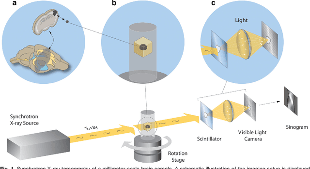
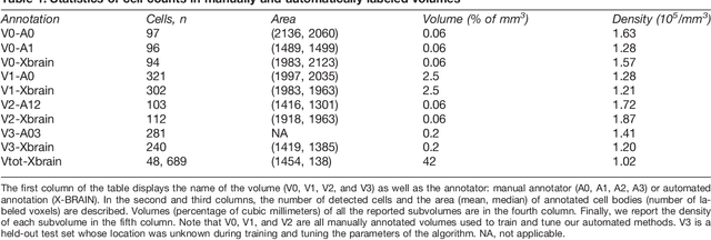
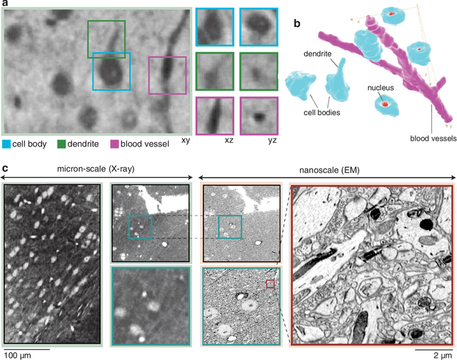
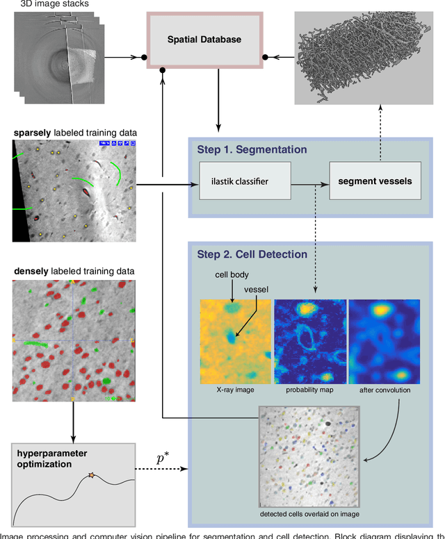
Methods for resolving the 3D microstructure of the brain typically start by thinly slicing and staining the brain, and then imaging each individual section with visible light photons or electrons. In contrast, X-rays can be used to image thick samples, providing a rapid approach for producing large 3D brain maps without sectioning. Here we demonstrate the use of synchrotron X-ray microtomography ($\mu$CT) for producing mesoscale $(1~\mu m^3)$ resolution brain maps from millimeter-scale volumes of mouse brain. We introduce a pipeline for $\mu$CT-based brain mapping that combines methods for sample preparation, imaging, automated segmentation of image volumes into cells and blood vessels, and statistical analysis of the resulting brain structures. Our results demonstrate that X-ray tomography promises rapid quantification of large brain volumes, complementing other brain mapping and connectomics efforts.