Jing W. Zhang
Deep learning model trained on mobile phone-acquired frozen section images effectively detects basal cell carcinoma
Nov 22, 2020


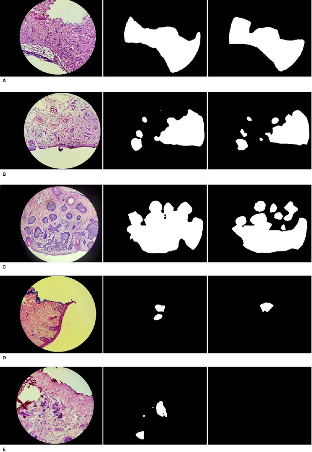
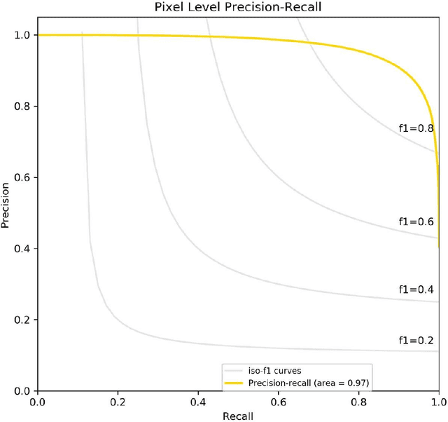
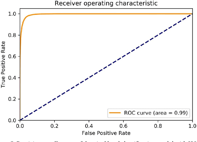
Abstract:Background: Margin assessment of basal cell carcinoma using the frozen section is a common task of pathology intraoperative consultation. Although frequently straight-forward, the determination of the presence or absence of basal cell carcinoma on the tissue sections can sometimes be challenging. We explore if a deep learning model trained on mobile phone-acquired frozen section images can have adequate performance for future deployment. Materials and Methods: One thousand two hundred and forty-one (1241) images of frozen sections performed for basal cell carcinoma margin status were acquired using mobile phones. The photos were taken at 100x magnification (10x objective). The images were downscaled from a 4032 x 3024 pixel resolution to 576 x 432 pixel resolution. Semantic segmentation algorithm Deeplab V3 with Xception backbone was used for model training. Results: The model uses an image as input and produces a 2-dimensional black and white output of prediction of the same dimension; the areas determined to be basal cell carcinoma were displayed with white color, in a black background. Any output with the number of white pixels exceeding 0.5% of the total number of pixels is deemed positive for basal cell carcinoma. On the test set, the model achieves area under curve of 0.99 for receiver operator curve and 0.97 for precision-recall curve at the pixel level. The accuracy of classification at the slide level is 96%. Conclusions: The deep learning model trained with mobile phone images shows satisfactory performance characteristics, and thus demonstrates the potential for deploying as a mobile phone app to assist in frozen section interpretation in real time.
 Add to Chrome
 Add to Firefox
 Add to Edge