Jean-Luc Descotes
TIMC
First Clinical Experience in Urologic Surgery with a Novel Robotic Lightweight Laparoscope Holder
Aug 31, 2012


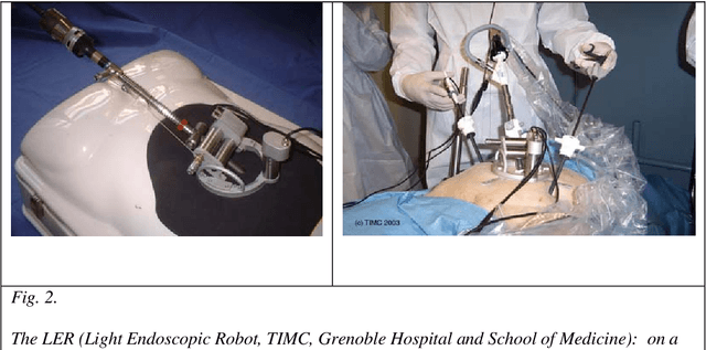
Abstract:Purpose: To report the feasibility and the safety of a surgeon-controlled robotic endoscope holder in laparoscopic surgery. Materials and methods: From March 2010 to September 2010, 20 patients were enrolled prospectively to undergo a laparoscopic surgery using an innovative robotic endoscope holder. Two surgeons performed 6 adrenalectomies, 4 sacrocolpopexies, 5 pyeloplasties, 4 radical prostatectomies and 1 radical nephrectomy. Demographic data, overall set-up time, operative time, number of assistants needed were reviewed. Surgeon's satisfaction regarding the ergonomics was assessed using a ten point scale. Postoperative clinical outcomes were reviewed at day 1 and 1 month postoperatively. Results: The per-protocol analysis was performed on 17 patients for whom the robot was effectively used for surgery. Median age was 63 years, 10 patients were female (59%). Median BMI was 26.8. Surgical procedures were completed with the robot in 12 cases (71 %). Median number of surgical assistant was 0. Overall set-up time with the robot was 19 min, operative time was 130 min) during which the robot was used 71% of the time. Mean hospital stay was 6.94 days $\pm$ 2.3. Median score regarding the easiness of use was 7. Median pain level was 1.5/10 at day 1 and 0 at 1 month postoperatively. Open conversion was needed in 1 case (6 %) and 4 minor complications occurred in 2 patients (12%). Conclusion: This use of this novel robotic laparoscope holder is safe, feasible and it provides a good comfort to the surgeon.
Development of a Novel Robot for Transperineal Needle Based Interventions: Focal Therapy, Brachytherapy and Prostate Biopsies
Aug 31, 2012


Abstract:Purpose: We report what is to our knowledge the initial experience with a new 3-dimensional ultrasound robotic system for prostate brachytherapy assistance, focal therapy and prostate biopsies. Its ability to track prostate motion intraoperatively allows it to manage motions and guide needles to predefined targets. Materials and Methods: A robotic system was created for transrectal ultrasound guided needle implantation combined with intraoperative prostate tracking. Experiments were done on 90 targets embedded in a total of 9 mobile, deformable, synthetic prostate phantoms. Experiments involved trying to insert glass beads as close as possible to targets in multimodal anthropomorphic imaging phantoms. Results were measured by segmenting the inserted beads in computerized tomography volumes of the phantoms. Results: The robot reached the chosen targets in phantoms with a median accuracy of 2.73 mm and a median prostate motion of 5.46 mm. Accuracy was better at the apex than at the base (2.28 vs 3.83 mm, p <0.001), and similar for horizontal and angled needle inclinations (2.7 vs 2.82 mm, p = 0.18). Conclusions: To our knowledge this robot for prostate focal therapy, brachytherapy and targeted prostate biopsies is the first system to use intraoperative prostate motion tracking to guide needles into the prostate. Preliminary experiments show its ability to reach targets despite prostate motion.
Prosper: image and robot-guided prostate brachytherapy
Apr 08, 2011

Abstract:Brachytherapy for localized prostate cancer consists in destroying cancer by introducing iodine radioactive seeds into the gland through hollow needles. The planning of the position of the seeds and their introduction into the prostate is based on intra-operative ultrasound (US) imaging. We propose to optimize the global quality of the procedure by: i) using 3D US; ii) enhancing US data with MRI registration; iii) using a specially designed needle-insertion robot, connected to the imaging data. The imaging methods have been successfully tested on patient data while the robot accuracy has been evaluated on a realistic deformable phantom.
Medical image computing and computer-aided medical interventions applied to soft tissues. Work in progress in urology
Dec 13, 2007


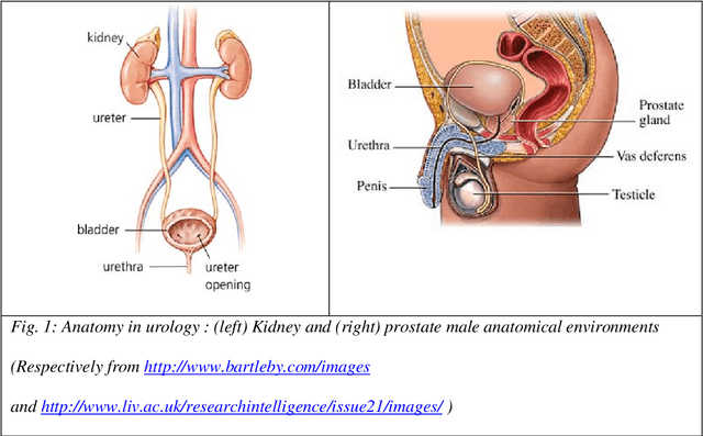
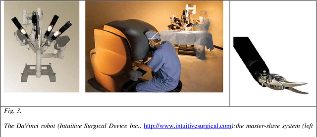
Abstract:Until recently, Computer-Aided Medical Interventions (CAMI) and Medical Robotics have focused on rigid and non deformable anatomical structures. Nowadays, special attention is paid to soft tissues, raising complex issues due to their mobility and deformation. Mini-invasive digestive surgery was probably one of the first fields where soft tissues were handled through the development of simulators, tracking of anatomical structures and specific assistance robots. However, other clinical domains, for instance urology, are concerned. Indeed, laparoscopic surgery, new tumour destruction techniques (e.g. HIFU, radiofrequency, or cryoablation), increasingly early detection of cancer, and use of interventional and diagnostic imaging modalities, recently opened new challenges to the urologist and scientists involved in CAMI. This resulted in the last five years in a very significant increase of research and developments of computer-aided urology systems. In this paper, we propose a description of the main problems related to computer-aided diagnostic and therapy of soft tissues and give a survey of the different types of assistance offered to the urologist: robotization, image fusion, surgical navigation. Both research projects and operational industrial systems are discussed.
 Add to Chrome
 Add to Firefox
 Add to Edge