Jane Garrison
Contrastive-Adversarial and Diffusion: Exploring pre-training and fine-tuning strategies for sulcal identification
May 29, 2024

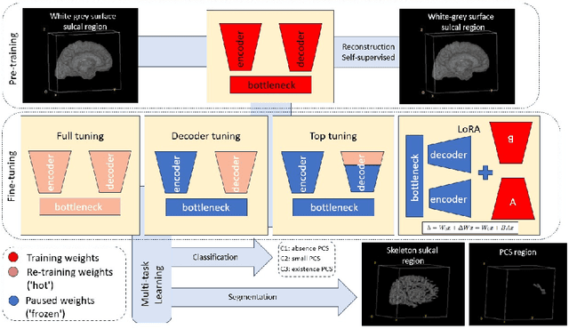
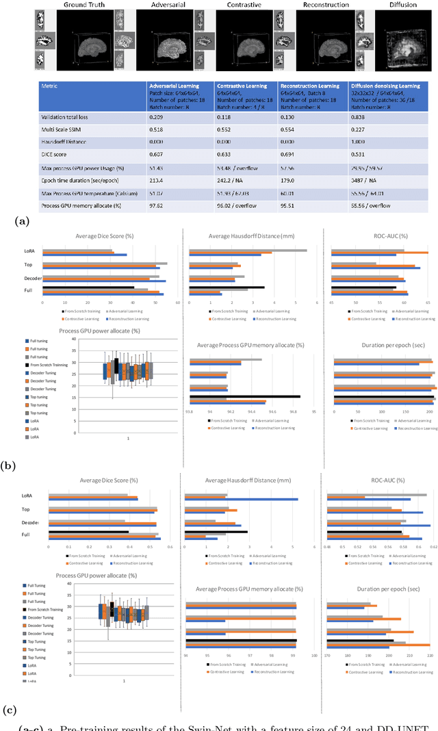
Abstract:In the last decade, computer vision has witnessed the establishment of various training and learning approaches. Techniques like adversarial learning, contrastive learning, diffusion denoising learning, and ordinary reconstruction learning have become standard, representing state-of-the-art methods extensively employed for fully training or pre-training networks across various vision tasks. The exploration of fine-tuning approaches has emerged as a current focal point, addressing the need for efficient model tuning with reduced GPU memory usage and time costs while enhancing overall performance, as exemplified by methodologies like low-rank adaptation (LoRA). Key questions arise: which pre-training technique yields optimal results - adversarial, contrastive, reconstruction, or diffusion denoising? How does the performance of these approaches vary as the complexity of fine-tuning is adjusted? This study aims to elucidate the advantages of pre-training techniques and fine-tuning strategies to enhance the learning process of neural networks in independent identical distribution (IID) cohorts. We underscore the significance of fine-tuning by examining various cases, including full tuning, decoder tuning, top-level tuning, and fine-tuning of linear parameters using LoRA. Systematic summaries of model performance and efficiency are presented, leveraging metrics such as accuracy, time cost, and memory efficiency. To empirically demonstrate our findings, we focus on a multi-task segmentation-classification challenge involving the paracingulate sulcus (PCS) using different 3D Convolutional Neural Network (CNN) architectures by using the TOP-OSLO cohort comprising 596 subjects.
A 3D explainability framework to uncover learning patterns and crucial sub-regions in variable sulci recognition
Sep 02, 2023Abstract:Precisely identifying sulcal features in brain MRI is made challenging by the variability of brain folding. This research introduces an innovative 3D explainability frame-work that validates outputs from deep learning networks in their ability to detect the paracingulate sulcus, an anatomical feature that may or may not be present on the frontal medial surface of the human brain. This study trained and tested two networks, amalgamating local explainability techniques GradCam and SHAP with a dimensionality reduction method. The explainability framework provided both localized and global explanations, along with accuracy of classification results, revealing pertinent sub-regions contributing to the decision process through a post-fusion transformation of explanatory and statistical features. Leveraging the TOP-OSLO dataset of MRI acquired from patients with schizophrenia, greater accuracies of paracingulate sulcus detection (presence or absence) were found in the left compared to right hemispheres with distinct, but extensive sub-regions contributing to each classification outcome. The study also inadvertently highlighted the critical role of an unbiased annotation protocol in maintaining network performance fairness. Our proposed method not only offers automated, impartial annotations of a variable sulcus but also provides insights into the broader anatomical variations associated with its presence throughout the brain. The adoption of this methodology holds promise for instigating further explorations and inquiries in the field of neuroscience.
 Add to Chrome
 Add to Firefox
 Add to Edge