Huseyin Uvet
An ensemble deep learning approach to detect tumors on Mohs micrographic surgery slides
Apr 07, 2025Abstract:Mohs micrographic surgery (MMS) is the gold standard technique for removing high risk nonmelanoma skin cancer however, intraoperative histopathological examination demands significant time, effort, and professionality. The objective of this study is to develop a deep learning model to detect basal cell carcinoma (BCC) and artifacts on Mohs slides. A total of 731 Mohs slides from 51 patients with BCCs were used in this study, with 91 containing tumor and 640 without tumor which was defined as non-tumor. The dataset was employed to train U-Net based models that segment tumor and non-tumor regions on the slides. The segmented patches were classified as tumor, or non-tumor to produce predictions for whole slide images (WSIs). For the segmentation phase, the deep learning model success was measured using a Dice score with 0.70 and 0.67 value, area under the curve (AUC) score with 0.98 and 0.96 for tumor and non-tumor, respectively. For the tumor classification, an AUC of 0.98 for patch-based detection, and AUC of 0.91 for slide-based detection was obtained on the test dataset. We present an AI system that can detect tumors and non-tumors in Mohs slides with high success. Deep learning can aid Mohs surgeons and dermatopathologists in making more accurate decisions.
Super-Resolution for Interferometric Imaging: Model Comparisons and Performance Analysis
Feb 21, 2025Abstract:This study investigates the application of Super-Resolution techniques in holographic microscopy to enhance quantitative phase imaging. An off-axis Mach-Zehnder interferometric setup was employed to capture interferograms. The study evaluates two Super-Resolution models, RCAN and Real-ESRGAN, for their effectiveness in reconstructing high-resolution interferograms from a microparticle-based dataset. The models were assessed using two primary approaches: image-based analysis for structural detail enhancement and morphological evaluation for maintaining sample integrity and phase map accuracy. The results demonstrate that RCAN achieves superior numerical precision, making it ideal for applications requiring highly accurate phase map reconstruction, while Real-ESRGAN enhances visual quality and structural coherence, making it suitable for visualization-focused applications. This study highlights the potential of Super-Resolution models in overcoming diffraction-imposed resolution limitations in holographic microscopy, opening the way for improved imaging techniques in biomedical diagnostics, materials science, and other high-precision fields.
Comprehensive Study on Lumbar Disc Segmentation Techniques Using MRI Data
Dec 25, 2024Abstract:Lumbar disk segmentation is essential for diagnosing and curing spinal disorders by enabling precise detection of disk boundaries in medical imaging. The advent of deep learning has resulted in the development of many segmentation methods, offering differing levels of accuracy and effectiveness. This study assesses the effectiveness of several sophisticated deep learning architectures, including ResUnext, Ef3 Net, UNet, and TransUNet, for lumbar disk segmentation, highlighting key metrics like as Pixel Accuracy, Mean Intersection over Union (Mean IoU), and Dice Coefficient. The findings indicate that ResUnext achieved the highest segmentation accuracy, with a Pixel Accuracy of 0.9492 and a Dice Coefficient of 0.8425, with TransUNet following closely after. Filtering techniques somewhat enhanced the performance of most models, particularly Dense UNet, improving stability and segmentation quality. The findings underscore the efficacy of these models in lumbar disk segmentation and highlight potential areas for improvement.
CNN Based Detection of Cardiovascular Diseases from ECG Images
Aug 29, 2024Abstract:This study develops a Convolutional Neural Network (CNN) model for detecting myocardial infarction (MI) from Electrocardiogram (ECG) images. The model, built using the InceptionV3 architecture and optimized through transfer learning, was trained using ECG data obtained from the Ch. Pervaiz Elahi Institute of Cardiology in Pakistan. The dataset includes ECG images representing four different cardiac conditions: myocardial infarction, abnormal heartbeat, history of myocardial infarction, and normal heart activity. The developed model successfully detects MI and other cardiovascular conditions with an accuracy of 93.27%. This study demonstrates that deep learning-based models can provide significant support to clinicians in the early detection and prevention of heart attacks.
Impact of velocity and impact angle on football shot accuracy during fundamental trainings
Feb 07, 2023


Abstract:The purpose of this research is to create a machine learning-based smart coaching approach for football that can replace manual analysis with real-time feedback for trainers. In-depth analysis of football player data by humans is time-consuming, error-prone, and requires a lot of effort. This exploratory study demonstrates the feasibility of using a machine learning algorithm to enhance the effectiveness of player monitoring and training. The suggested approach uses machine learning to generate analytical insights and enable long-term monitoring of player performance. In the future, machine learning could use this technique to offer constructive criticism of football players. The system incorporates a homemade ball-throwing mechanism capable of launching the ball in a variety of directions and at varying velocities. The ball kicker is equipped with a gyroscope and accelerometer sensors for measuring velocity and acceleration. The gathered data is filtered initially, and then the data that has been processed is fed into the machine-learning algorithm. The algorithm will be trained on player performance data and will be able to provide real-time feedback to coaches on player performance and potential areas for improvement. Additionally, the system will be able to track player progress over time and provide coaches with a comprehensive view of player development. The ultimate goal is to improve player performance and reduce the workload for coaches by automating the analysis process.
Benchmarking of Lightweight Deep Learning Architectures for Skin Cancer Classification using ISIC 2017 Dataset
Oct 23, 2021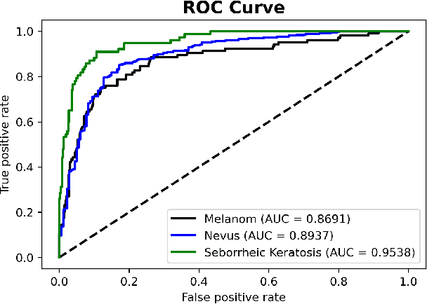


Abstract:Skin cancer is one of the deadly types of cancer and is common in the world. Recently, there has been a huge jump in the rate of people getting skin cancer. For this reason, the number of studies on skin cancer classification with deep learning are increasing day by day. For the growth of work in this area, the International Skin Imaging Collaboration (ISIC) organization was established and they created an open dataset archive. In this study, images were taken from ISIC 2017 Challenge. The skin cancer images taken were preprocessed and data augmented. Later, these images were trained with transfer learning and fine-tuning approach and deep learning models were created in this way. 3 different mobile deep learning models and 3 different batch size values were determined for each, and a total of 9 models were created. Among these models, the NASNetMobile model with 16 batch size got the best result. The accuracy value of this model is 82.00%, the precision value is 81.77% and the F1 score value is 0.8038. Our method is to benchmark mobile deep learning models which have few parameters and compare the results of the models.
Mechatronic Investigation of Wound Healing Process by Using Micro Robot
Aug 05, 2021


Abstract:The purpose of this study is to find ideal forces for reducing cell stress in wound healing process by micro robots. Because of this aim, we made two simulations on COMSOL Multiphysics with micro robot to find correct force. As a result of these simulation, we created force curves to obtain the minimum force and friction force that could lift the cells from the surface will be determined. As the potential of the system for two micro robots that have 2 mm x 0.25 mm x 0.4 mm dimension SU-8 body with 3 NdFeB that have 0.25 thickness and diameter, simulation results at maximum force in the x-axis calculated with 4.640 mN, the distance between the two robots is 150 um.
Automated Onychomycosis Detection Using Deep Neural Networks
Jul 13, 2021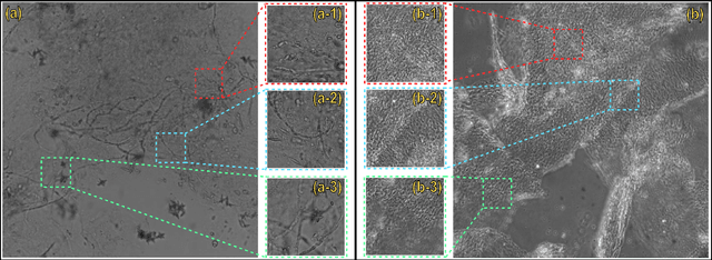
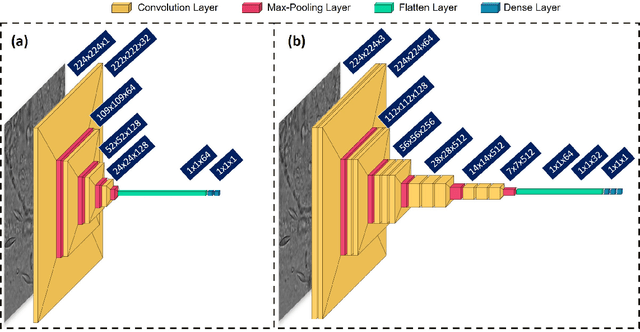
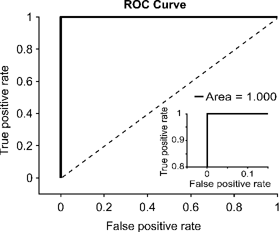

Abstract:Clinical dermatology, still relies heavily on manual introspection of fungi within a Potassium Hydroxide (KOH) solution using a brightfield microscope. However, this method takes a long time, is based on the experience of the clinician, and has a low accuracy. With the increase of neural network applications in the field of clinical microscopy it is now possible to automate such manual processes increasing both efficiency and accuracy. This study presents a deep neural network structure that enables the rapid solutions for these problems and can perform automatic fungi detection in grayscale images without colorants. Microscopic images of 81 fungi and 235 ceratine were collected. Then, smaller patches were extracted containing 2062 fungi and 2142 ceratine. In order to detect fungus and ceratine, two models were created one of which was a custom neural network and the other was based on the VGG16 architecture. The developed custom model had 99.84% accuracy, and an area under the curve (AUC) value of 1.00, while the VGG16 model had 98.89% accuracy and an AUC value of 0.99. However, average accuracy and AUC value of clinicians is 72.8% and 0.87 respectively. This deep learning model allows the development of an automated system that can detect fungi within microscopic images.
The Effect of Pore Structure in Flapping Wings on Flight Performance
Jun 03, 2021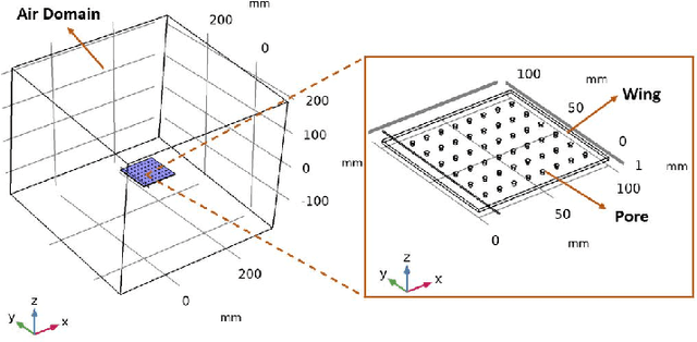
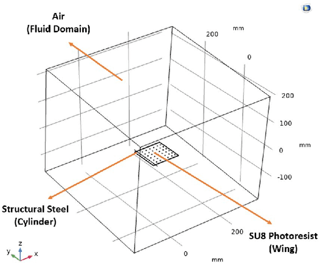


Abstract:This study investigates the effects of porosity on flying creatures such as dragonflies, moths, hummingbirds, etc. wing and shows that pores can affect wing performance. These studies were performed by 3D porous flapping wing flow analyses on Comsol Multiphysics. In this study, we analyzed different numbers of the porous wing at different angles of inclination in order to see the effect of pores on lift and drag forces. To compare the results 9 different analyses were performed. In these analyses, airflow velocity was taken as 5 m/s, angle of attack as 5 degrees, frequency as 25 Hz, and flapping angle as 30 degrees. By keeping these values constant, the number of pores was changed to 36, 48, and 60, and the pore angles of inclination to 60, 70, and 80 degrees. Analyses were carried out by giving laminar flow to this wing designed in the Comsol Multiphysics program. The importance of pores was investigated by comparing the results of these analyses.
Holographic Cell Stiffness Mapping Using Acoustic Stimulation
Feb 15, 2021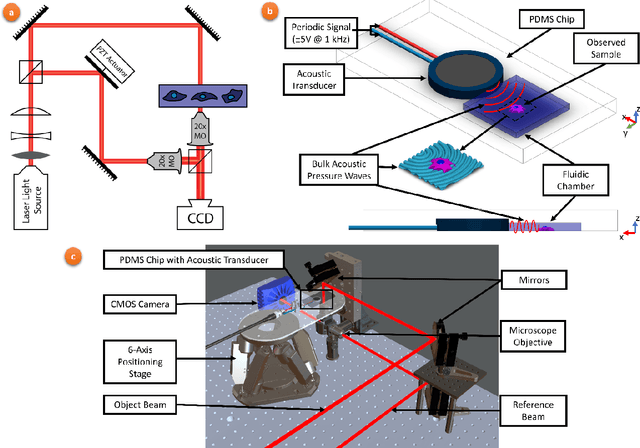
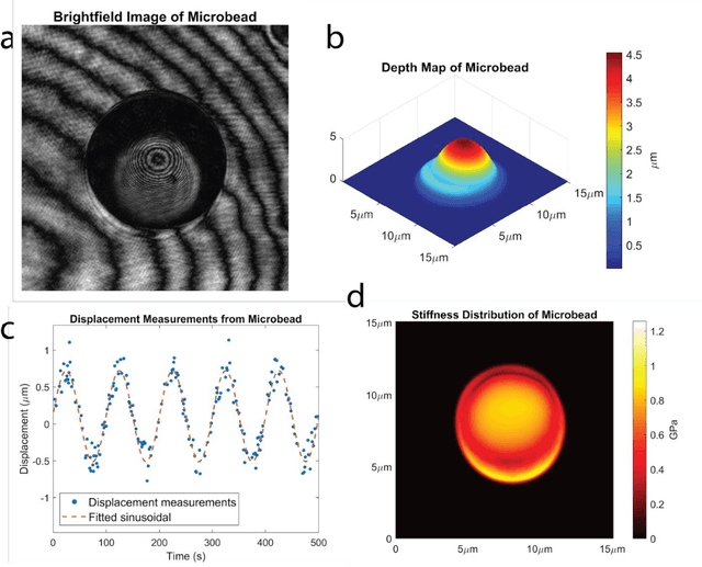

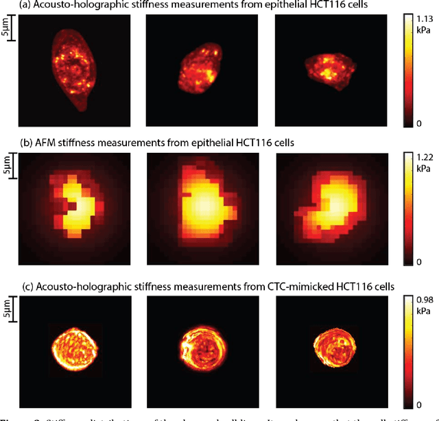
Abstract:Accurate assessment of stiffness distribution is essential due to the critical role of single cell mechanobiology in the regulation of many vital cellular processes such as proliferation, adhesion, migration, and motility. Cell stiffness is one of the fundamental mechanical properties of the cell and is greatly affected by the intracellular tensional forces, cytoskeletal prestress, and cytoskeleton structure. Herein, we propose a novel holographic single-cell stiffness measurement technique that can obtain the stiffness distribution over a cell membrane at high resolution and in real-time. The proposed imaging method coupled with acoustic signals allows us to assess the cell stiffness distribution with a low error margin and label-free manner. We demonstrate the proposed technique on HCT116 (Human Colorectal Carcinoma) cells and CTC-mimicked HCT116 cells by induction with transforming growth factor-beta (TGF-\b{eta}). Validation studies of the proposed approach were carried out on certified polystyrene microbeads with known stiffness levels. Its performance was evaluated in comparison with the AFM results obtained for the relevant cells. When the experimental results were examined, the proposed methodology shows utmost performance over average cell stiffness values for HCT116, and CTC-mimicked HCT116 cells were found as 1.08 kPa, and 0.88 kPa, respectively. The results confirm that CTC-mimicked HCT116 cells lose their adhesion ability to enter the vascular circulation and metastasize. They also exhibit a softer stiffness profile compared to adherent forms of the cancer cells. Hence, the proposed technique is a significant, reliable, and faster alternative for in-vitro cell stiffness characterization tools. It can be utilized for various applications where single-cell analysis is required, such as disease modeling, drug testing, diagnostics, and many more.
 Add to Chrome
 Add to Firefox
 Add to Edge