Fernando Vega
Towards Integrating Epistemic Uncertainty Estimation into the Radiotherapy Workflow
Sep 27, 2024Abstract:The precision of contouring target structures and organs-at-risk (OAR) in radiotherapy planning is crucial for ensuring treatment efficacy and patient safety. Recent advancements in deep learning (DL) have significantly improved OAR contouring performance, yet the reliability of these models, especially in the presence of out-of-distribution (OOD) scenarios, remains a concern in clinical settings. This application study explores the integration of epistemic uncertainty estimation within the OAR contouring workflow to enable OOD detection in clinically relevant scenarios, using specifically compiled data. Furthermore, we introduce an advanced statistical method for OOD detection to enhance the methodological framework of uncertainty estimation. Our empirical evaluation demonstrates that epistemic uncertainty estimation is effective in identifying instances where model predictions are unreliable and may require an expert review. Notably, our approach achieves an AUC-ROC of 0.95 for OOD detection, with a specificity of 0.95 and a sensitivity of 0.92 for implant cases, underscoring its efficacy. This study addresses significant gaps in the current research landscape, such as the lack of ground truth for uncertainty estimation and limited empirical evaluations. Additionally, it provides a clinically relevant application of epistemic uncertainty estimation in an FDA-approved and widely used clinical solution for OAR segmentation from Varian, a Siemens Healthineers company, highlighting its practical benefits.
Three-Dimensional Amyloid-Beta PET Synthesis from Structural MRI with Conditional Generative Adversarial Networks
May 03, 2024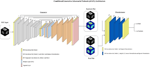
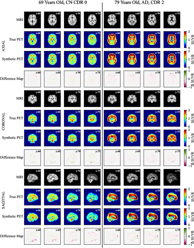
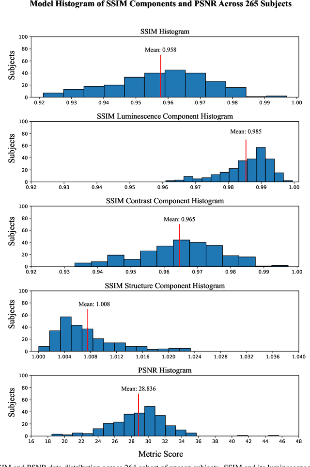

Abstract:Motivation: Alzheimer's Disease hallmarks include amyloid-beta deposits and brain atrophy, detectable via PET and MRI scans, respectively. PET is expensive, invasive and exposes patients to ionizing radiation. MRI is cheaper, non-invasive, and free from ionizing radiation but limited to measuring brain atrophy. Goal: To develop an 3D image translation model that synthesizes amyloid-beta PET images from T1-weighted MRI, exploiting the known relationship between amyloid-beta and brain atrophy. Approach: The model was trained on 616 PET/MRI pairs and validated with 264 pairs. Results: The model synthesized amyloid-beta PET images from T1-weighted MRI with high-degree of similarity showing high SSIM and PSNR metrics (SSIM>0.95&PSNR=28). Impact: Our model proves the feasibility of synthesizing amyloid-beta PET images from structural MRI ones, significantly enhancing accessibility for large-cohort studies and early dementia detection, while also reducing cost, invasiveness, and radiation exposure.
Machine Learning-based Estimation of Respiratory Fluctuations in a Healthy Adult Population using BOLD fMRI and Head Motion Parameters
Apr 30, 2024


Abstract:Motivation: In many fMRI studies, respiratory signals are often missing or of poor quality. Therefore, it could be highly beneficial to have a tool to extract respiratory variation (RV) waveforms directly from fMRI data without the need for peripheral recording devices. Goal(s): Investigate the hypothesis that head motion parameters contain valuable information regarding respiratory patter, which can help machine learning algorithms estimate the RV waveform. Approach: This study proposes a CNN model for reconstruction of RV waveforms using head motion parameters and BOLD signals. Results: This study showed that combining head motion parameters with BOLD signals enhances RV waveform estimation. Impact: It is expected that application of the proposed method will lower the cost of fMRI studies, reduce complexity, and decrease the burden on participants as they will not be required to wear a respiratory bellows.
A voxel-level approach to brain age prediction: A method to assess regional brain aging
Oct 17, 2023


Abstract:Brain aging is a regional phenomenon, a facet that remains relatively under-explored within the realm of brain age prediction research using machine learning methods. Voxel-level predictions can provide localized brain age estimates that can provide granular insights into the regional aging processes. This is essential to understand the differences in aging trajectories in healthy versus diseased subjects. In this work, a deep learning-based multitask model is proposed for voxel-level brain age prediction from T1-weighted magnetic resonance images. The proposed model outperforms the models existing in the literature and yields valuable clinical insights when applied to both healthy and diseased populations. Regional analysis is performed on the voxel-level brain age predictions to understand aging trajectories of known anatomical regions in the brain and show that there exist disparities in regional aging trajectories of healthy subjects compared to ones with underlying neurological disorders such as Dementia and more specifically, Alzheimer's disease. Our code is available at https://github.com/nehagianchandani/Voxel-level-brain-age-prediction.
Amyloid-Beta Axial Plane PET Synthesis from Structural MRI: An Image Translation Approach for Screening Alzheimer's Disease
Sep 01, 2023
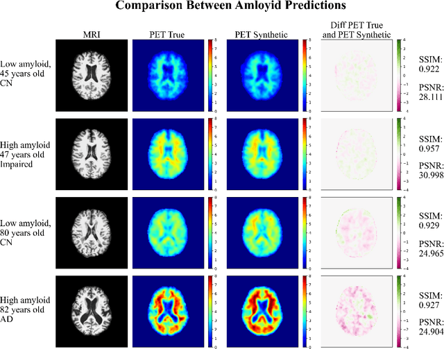

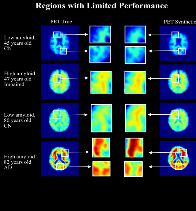
Abstract:In this work, an image translation model is implemented to produce synthetic amyloid-beta PET images from structural MRI that are quantitatively accurate. Image pairs of amyloid-beta PET and structural MRI were used to train the model. We found that the synthetic PET images could be produced with a high degree of similarity to truth in terms of shape, contrast and overall high SSIM and PSNR. This work demonstrates that performing structural to quantitative image translation is feasible to enable the access amyloid-beta information from only MRI.
Denoising Simulated Low-Field MRI (70mT) using Denoising Autoencoders (DAE) and Cycle-Consistent Generative Adversarial Networks (Cycle-GAN)
Jul 12, 2023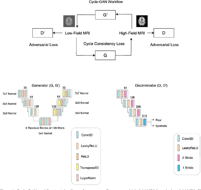
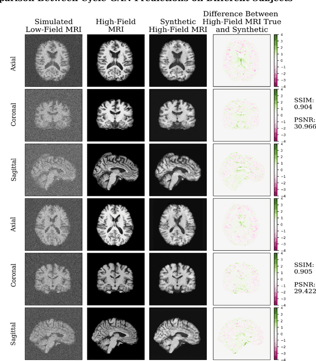
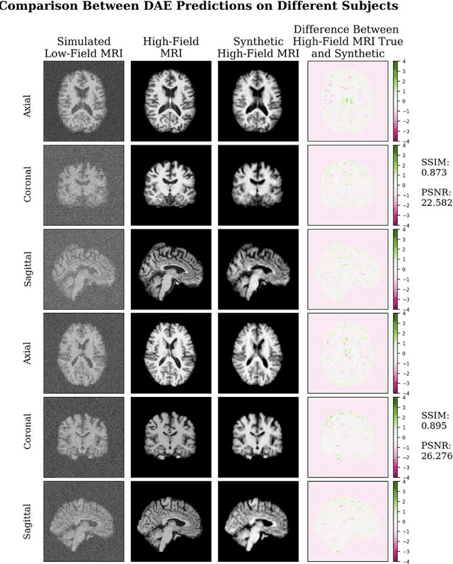
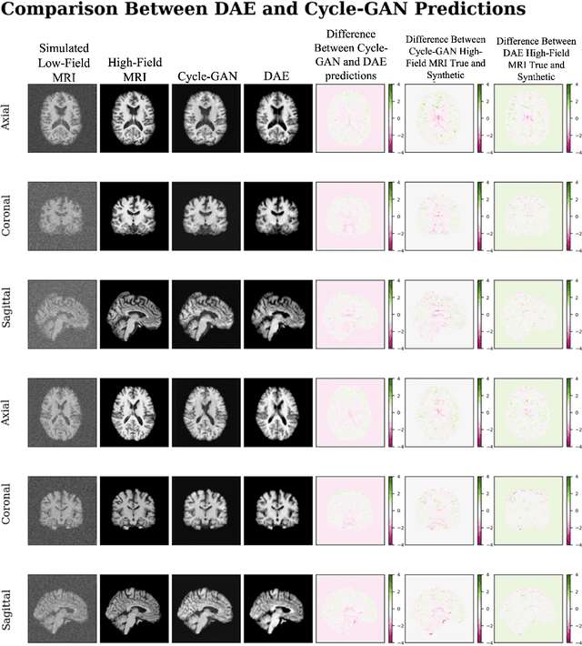
Abstract:In this work, a denoising Cycle-GAN (Cycle Consistent Generative Adversarial Network) is implemented to yield high-field, high resolution, high signal-to-noise ratio (SNR) Magnetic Resonance Imaging (MRI) images from simulated low-field, low resolution, low SNR MRI images. Resampling and additive Rician noise were used to simulate low-field MRI. Images were utilized to train a Denoising Autoencoder (DAE) and a Cycle-GAN, with paired and unpaired cases. Both networks were evaluated using SSIM and PSNR image quality metrics. This work demonstrates the use of a generative deep learning model that can outperform classical DAEs to improve low-field MRI images and does not require image pairs.
Using BOLD-fMRI to Compute the Respiration Volume per Time and Respiration Variation with Convolutional Neural Networks in the Human Connectome Development Cohort
Jul 03, 2023


Abstract:In many fMRI studies, respiratory signals are unavailable or do not have acceptable quality. Consequently, the direct removal of low-frequency respiratory variations from BOLD signals is not possible. This study proposes a one-dimensional CNN model for reconstruction of two respiratory measures, RV and RVT. Results show that a CNN can capture informative features from resting BOLD signals and reconstruct realistic RV and RVT timeseries. It is expected that application of the proposed method will lower the cost of fMRI studies, reduce complexity, and decrease the burden on participants as they will not be required to wear a respiratory bellows.
 Add to Chrome
 Add to Firefox
 Add to Edge