Fabian Laumer
From Slices to Structures: Unsupervised 3D Reconstruction of Female Pelvic Anatomy from Freehand Transvaginal Ultrasound
Aug 20, 2025Abstract:Volumetric ultrasound has the potential to significantly improve diagnostic accuracy and clinical decision-making, yet its widespread adoption remains limited by dependence on specialized hardware and restrictive acquisition protocols. In this work, we present a novel unsupervised framework for reconstructing 3D anatomical structures from freehand 2D transvaginal ultrasound (TVS) sweeps, without requiring external tracking or learned pose estimators. Our method adapts the principles of Gaussian Splatting to the domain of ultrasound, introducing a slice-aware, differentiable rasterizer tailored to the unique physics and geometry of ultrasound imaging. We model anatomy as a collection of anisotropic 3D Gaussians and optimize their parameters directly from image-level supervision, leveraging sensorless probe motion estimation and domain-specific geometric priors. The result is a compact, flexible, and memory-efficient volumetric representation that captures anatomical detail with high spatial fidelity. This work demonstrates that accurate 3D reconstruction from 2D ultrasound images can be achieved through purely computational means, offering a scalable alternative to conventional 3D systems and enabling new opportunities for AI-assisted analysis and diagnosis.
Interpretable Anomaly Detection in Echocardiograms with Dynamic Variational Trajectory Models
Jun 30, 2022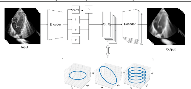
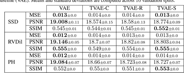
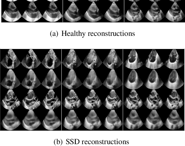

Abstract:We propose a novel anomaly detection method for echocardiogram videos. The introduced method takes advantage of the periodic nature of the heart cycle to learn different variants of a variational latent trajectory model (TVAE). The models are trained on the healthy samples of an in-house dataset of infant echocardiogram videos consisting of multiple chamber views to learn a normative prior of the healthy population. During inference, maximum a posteriori (MAP) based anomaly detection is performed to detect out-of-distribution samples in our dataset. The proposed method reliably identifies severe congenital heart defects, such as Ebstein's Anomaly or Shonecomplex. Moreover, it achieves superior performance over MAP-based anomaly detection with standard variational autoencoders on the task of detecting pulmonary hypertension and right ventricular dilation. Finally, we demonstrate that the proposed method provides interpretable explanations of its output through heatmaps which highlight the regions corresponding to anomalous heart structures.
Interpretable Prediction of Pulmonary Hypertension in Newborns using Echocardiograms
Mar 24, 2022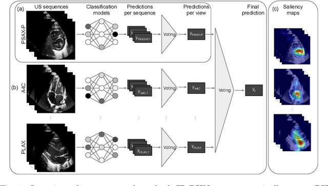
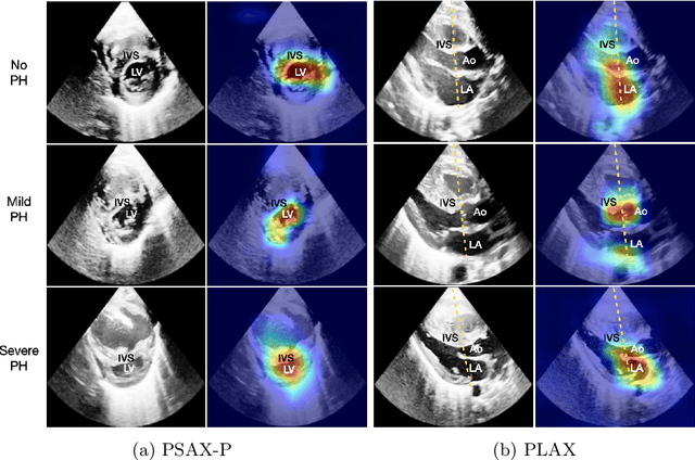
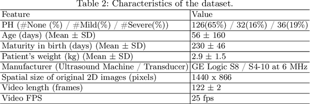
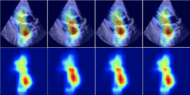
Abstract:Pulmonary hypertension (PH) in newborns and infants is a complex condition associated with several pulmonary, cardiac, and systemic diseases contributing to morbidity and mortality. Therefore, accurate and early detection of PH is crucial for successful management. Using echocardiography, the primary diagnostic tool in pediatrics, human assessment is both time-consuming and expertise-demanding, raising the need for an automated approach. In this work, we present an interpretable multi-view video-based deep learning approach to predict PH for a cohort of 194 newborns using echocardiograms. We use spatio-temporal convolutional architectures for the prediction of PH from each view, and aggregate the predictions of the different views using majority voting. To the best of our knowledge, this is the first work for an automated assessment of PH in newborns using echocardiograms. Our results show a mean F1-score of 0.84 for severity prediction and 0.92 for binary detection using 10-fold cross-validation. We complement our predictions with saliency maps and show that the learned model focuses on clinically relevant cardiac structures, motivating its usage in clinical practice.
Neural collaborative filtering for unsupervised mitral valve segmentation in echocardiography
Aug 13, 2020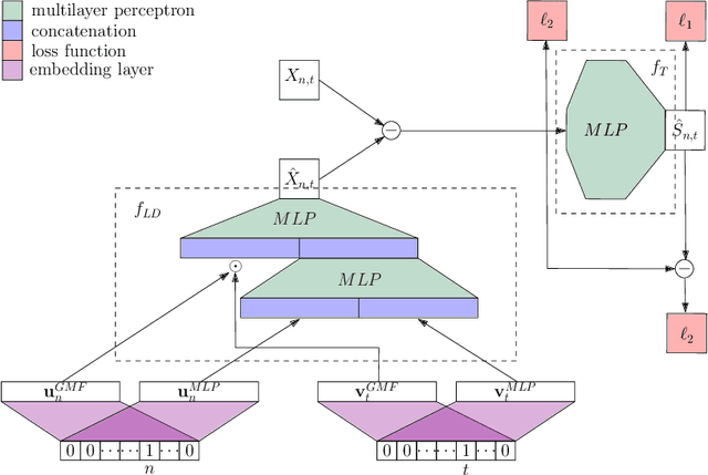
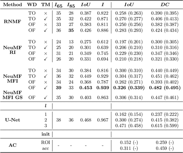
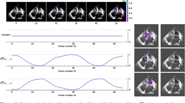
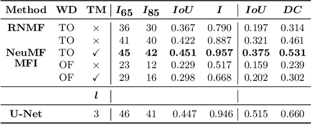
Abstract:The segmentation of the mitral valve annulus and leaflets specifies a crucial first step to establish a machine learning pipeline that can support physicians in performing multiple tasks, e.g.\ diagnosis of mitral valve diseases, surgical planning, and intraoperative procedures. Current methods for mitral valve segmentation on 2D echocardiography videos require extensive interaction with annotators and perform poorly on low-quality and noisy videos. We propose an automated and unsupervised method for the mitral valve segmentation based on a low dimensional embedding of the echocardiography videos using neural network collaborative filtering. The method is evaluated in a collection of echocardiography videos of patients with a variety of mitral valve diseases, and additionally on an independent test cohort. It outperforms state-of-the-art \emph{unsupervised} and \emph{supervised} methods on low-quality videos or in the case of sparse annotation.
 Add to Chrome
 Add to Firefox
 Add to Edge