Elizabeth J. Atkinson
A Deep Learning-Based Method for Automatic Segmentation of Proximal Femur from Quantitative Computed Tomography Images
Jul 01, 2020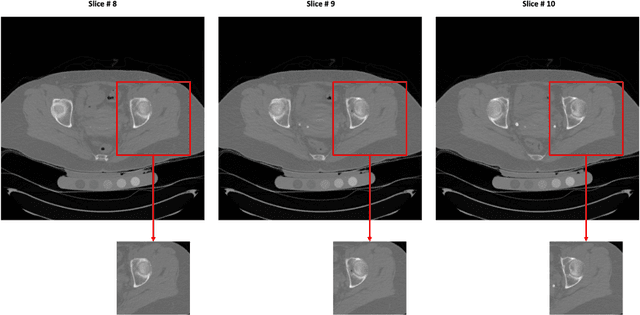
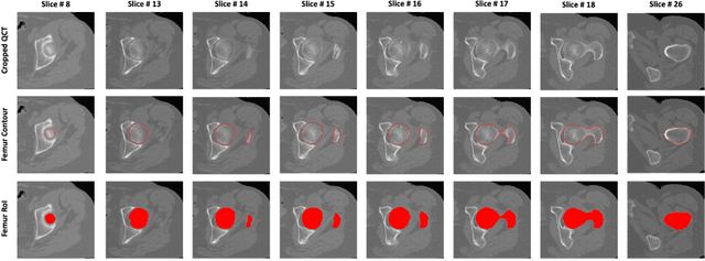

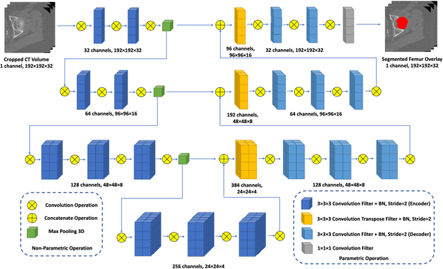
Abstract:Purpose: Proximal femur image analyses based on quantitative computed tomography (QCT) provide a method to quantify the bone density and evaluate osteoporosis and risk of fracture. We aim to develop a deep-learning-based method for automatic proximal femur segmentation. Methods and Materials: We developed a 3D image segmentation method based on V-Net, an end-to-end fully convolutional neural network (CNN), to extract the proximal femur QCT images automatically. The proposed V-net methodology adopts a compound loss function, which includes a Dice loss and a L2 regularizer. We performed experiments to evaluate the effectiveness of the proposed segmentation method. In the experiments, a QCT dataset which included 397 QCT subjects was used. For the QCT image of each subject, the ground truth for the proximal femur was delineated by a well-trained scientist. During the experiments for the entire cohort then for male and female subjects separately, 90% of the subjects were used in 10-fold cross-validation for training and internal validation, and to select the optimal parameters of the proposed models; the rest of the subjects were used to evaluate the performance of models. Results: Visual comparison demonstrated high agreement between the model prediction and ground truth contours of the proximal femur portion of the QCT images. In the entire cohort, the proposed model achieved a Dice score of 0.9815, a sensitivity of 0.9852 and a specificity of 0.9992. In addition, an R2 score of 0.9956 (p<0.001) was obtained when comparing the volumes measured by our model prediction with the ground truth. Conclusion: This method shows a great promise for clinical application to QCT and QCT-based finite element analysis of the proximal femur for evaluating osteoporosis and hip fracture risk.
Unsupervised Machine Learning for the Discovery of Latent Disease Clusters and Patient Subgroups Using Electronic Health Records
May 17, 2019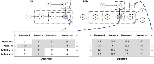

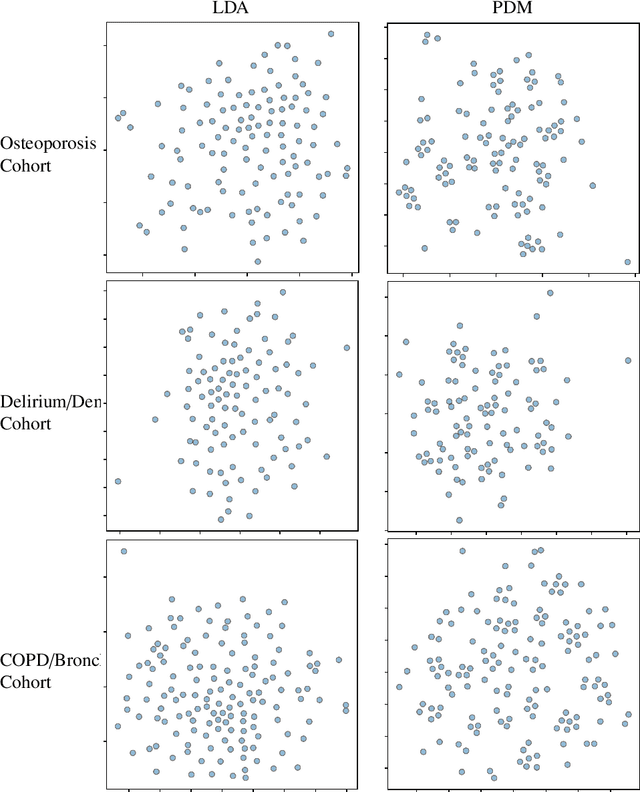
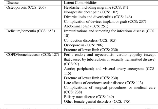
Abstract:Machine learning has become ubiquitous and a key technology on mining electronic health records (EHRs) for facilitating clinical research and practice. Unsupervised machine learning, as opposed to supervised learning, has shown promise in identifying novel patterns and relations from EHRs without using human created labels. In this paper, we investigate the application of unsupervised machine learning models in discovering latent disease clusters and patient subgroups based on EHRs. We utilized Latent Dirichlet Allocation (LDA), a generative probabilistic model, and proposed a novel model named Poisson Dirichlet Model (PDM), which extends the LDA approach using a Poisson distribution to model patients' disease diagnoses and to alleviate age and sex factors by considering both observed and expected observations. In the empirical experiments, we evaluated LDA and PDM on three patient cohorts with EHR data retrieved from the Rochester Epidemiology Project (REP), for the discovery of latent disease clusters and patient subgroups. We compared the effectiveness of LDA and PDM in identifying latent disease clusters through the visualization of disease representations learned by two approaches. We also tested the performance of LDA and PDM in differentiating patient subgroups through survival analysis, as well as statistical analysis. The experimental results show that the proposed PDM could effectively identify distinguished disease clusters by alleviating the impact of age and sex, and that LDA could stratify patients into more differentiable subgroups than PDM in terms of p-values. However, the subgroups discovered by PDM might imply the underlying patterns of diseases of greater interest in epidemiology research due to the alleviation of age and sex. Both unsupervised machine learning approaches could be leveraged to discover patient subgroups using EHRs but with different foci.
 Add to Chrome
 Add to Firefox
 Add to Edge