Bingchun Luo
MIPI 2024 Challenge on Demosaic for HybridEVS Camera: Methods and Results
May 08, 2024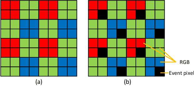
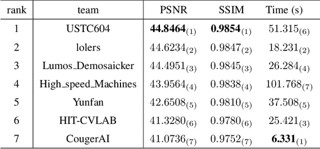


Abstract:The increasing demand for computational photography and imaging on mobile platforms has led to the widespread development and integration of advanced image sensors with novel algorithms in camera systems. However, the scarcity of high-quality data for research and the rare opportunity for in-depth exchange of views from industry and academia constrain the development of mobile intelligent photography and imaging (MIPI). Building on the achievements of the previous MIPI Workshops held at ECCV 2022 and CVPR 2023, we introduce our third MIPI challenge including three tracks focusing on novel image sensors and imaging algorithms. In this paper, we summarize and review the Nighttime Flare Removal track on MIPI 2024. In total, 170 participants were successfully registered, and 14 teams submitted results in the final testing phase. The developed solutions in this challenge achieved state-of-the-art performance on Nighttime Flare Removal. More details of this challenge and the link to the dataset can be found at https://mipi-challenge.org/MIPI2024/.
US-SFNet: A Spatial-Frequency Domain-based Multi-branch Network for Cervical Lymph Node Lesions Diagnoses in Ultrasound Images
Aug 31, 2023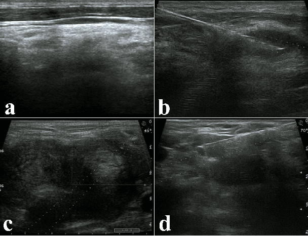
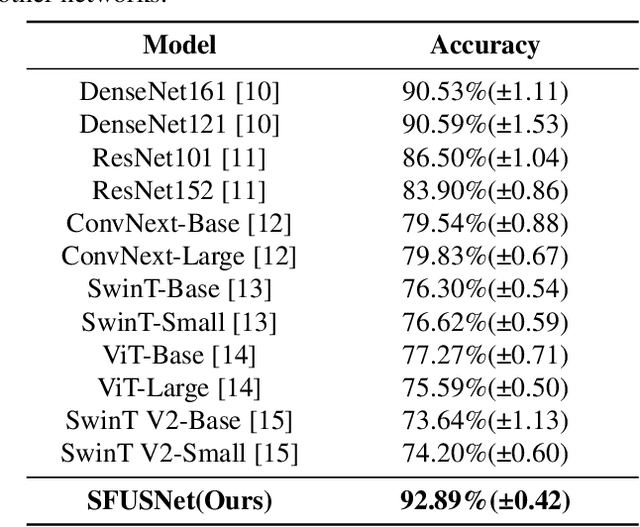
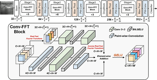
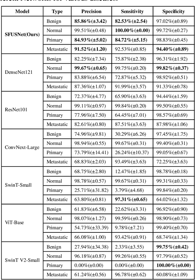
Abstract:Ultrasound imaging serves as a pivotal tool for diagnosing cervical lymph node lesions. However, the diagnoses of these images largely hinge on the expertise of medical practitioners, rendering the process susceptible to misdiagnoses. Although rapidly developing deep learning has substantially improved the diagnoses of diverse ultrasound images, there remains a conspicuous research gap concerning cervical lymph nodes. The objective of our work is to accurately diagnose cervical lymph node lesions by leveraging a deep learning model. To this end, we first collected 3392 images containing normal lymph nodes, benign lymph node lesions, malignant primary lymph node lesions, and malignant metastatic lymph node lesions. Given that ultrasound images are generated by the reflection and scattering of sound waves across varied bodily tissues, we proposed the Conv-FFT Block. It integrates convolutional operations with the fast Fourier transform to more astutely model the images. Building upon this foundation, we designed a novel architecture, named US-SFNet. This architecture not only discerns variances in ultrasound images from the spatial domain but also adeptly captures microstructural alterations across various lesions in the frequency domain. To ascertain the potential of US-SFNet, we benchmarked it against 12 popular architectures through five-fold cross-validation. The results show that US-SFNet is SOTA and can achieve 92.89% accuracy, 90.46% precision, 89.95% sensitivity and 97.49% specificity, respectively.
 Add to Chrome
 Add to Firefox
 Add to Edge