Haihua Liang
U-SEANNet: A Simple, Efficient and Applied U-Shaped Network for Diagnosis of Nasal Diseases on Nasal Endoscopic Images
Sep 02, 2023Abstract:Numerous studies have affirmed that deep learning models can facilitate early diagnosis of lesions in endoscopic images. However, the lack of available datasets stymies advancements in research on nasal endoscopy, and existing models fail to strike a good trade-off between model diagnosis performance, model complexity and parameters size, rendering them unsuitable for real-world application. To bridge these gaps, we created the first large-scale nasal endoscopy dataset, named 7-NasalEID, comprising 11,352 images that contain six common nasal diseases and normal samples. Subsequently, we proposed U-SEANNet, an innovative U-shaped architecture, underpinned by depth-wise separable convolution. Moreover, to enhance its capacity for detecting nuanced discrepancies in input images, U-SEANNet employs the Global-Local Channel Feature Fusion module, enabling it to utilize salient channel features from both global and local contexts. To demonstrate U-SEANNet's potential, we benchmarked U-SEANNet against seventeen modern architectures through five-fold cross-validation. The experimental results show that U-SEANNet achieves a commendable accuracy of 93.58%. Notably, U-SEANNet's parameters size and GFLOPs are only 0.78M and 0.21, respectively. Our findings suggest U-SEANNet is the state-of-the-art model for nasal diseases diagnosis in endoscopic images.
US-SFNet: A Spatial-Frequency Domain-based Multi-branch Network for Cervical Lymph Node Lesions Diagnoses in Ultrasound Images
Aug 31, 2023


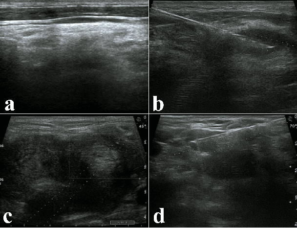
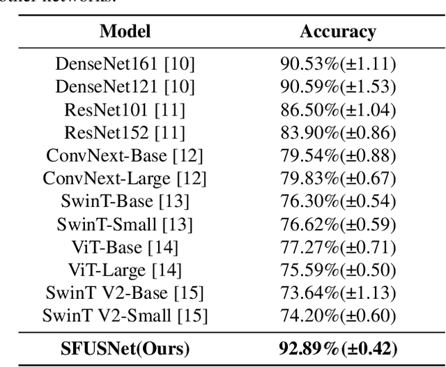
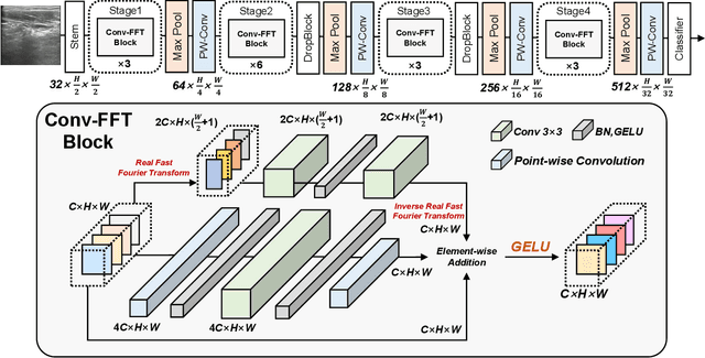
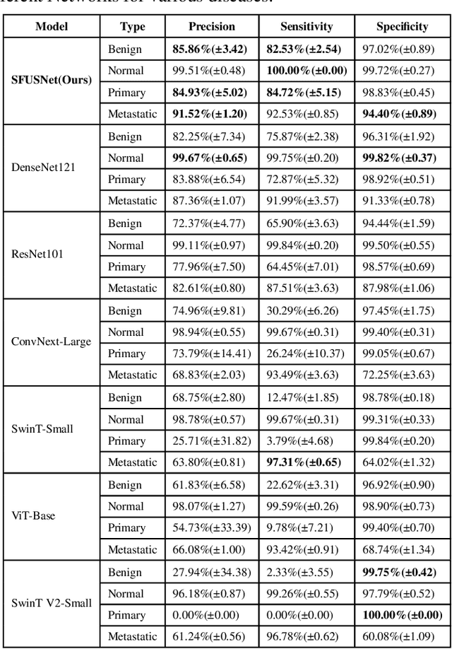
Abstract:Ultrasound imaging serves as a pivotal tool for diagnosing cervical lymph node lesions. However, the diagnoses of these images largely hinge on the expertise of medical practitioners, rendering the process susceptible to misdiagnoses. Although rapidly developing deep learning has substantially improved the diagnoses of diverse ultrasound images, there remains a conspicuous research gap concerning cervical lymph nodes. The objective of our work is to accurately diagnose cervical lymph node lesions by leveraging a deep learning model. To this end, we first collected 3392 images containing normal lymph nodes, benign lymph node lesions, malignant primary lymph node lesions, and malignant metastatic lymph node lesions. Given that ultrasound images are generated by the reflection and scattering of sound waves across varied bodily tissues, we proposed the Conv-FFT Block. It integrates convolutional operations with the fast Fourier transform to more astutely model the images. Building upon this foundation, we designed a novel architecture, named US-SFNet. This architecture not only discerns variances in ultrasound images from the spatial domain but also adeptly captures microstructural alterations across various lesions in the frequency domain. To ascertain the potential of US-SFNet, we benchmarked it against 12 popular architectures through five-fold cross-validation. The results show that US-SFNet is SOTA and can achieve 92.89% accuracy, 90.46% precision, 89.95% sensitivity and 97.49% specificity, respectively.
Ultrafast and Ultralight Network-Based Intelligent System for Real-time Diagnosis of Ear diseases in Any Devices
Aug 21, 2023


Abstract:Traditional ear disease diagnosis heavily depends on experienced specialists and specialized equipment, frequently resulting in misdiagnoses, treatment delays, and financial burdens for some patients. Utilizing deep learning models for efficient ear disease diagnosis has proven effective and affordable. However, existing research overlooked model inference speed and parameter size required for deployment. To tackle these challenges, we constructed a large-scale dataset comprising eight ear disease categories and normal ear canal samples from two hospitals. Inspired by ShuffleNetV2, we developed Best-EarNet, an ultrafast and ultralight network enabling real-time ear disease diagnosis. Best-EarNet incorporates the novel Local-Global Spatial Feature Fusion Module which can capture global and local spatial information simultaneously and guide the network to focus on crucial regions within feature maps at various levels, mitigating low accuracy issues. Moreover, our network uses multiple auxiliary classification heads for efficient parameter optimization. With 0.77M parameters, Best-EarNet achieves an average frames per second of 80 on CPU. Employing transfer learning and five-fold cross-validation with 22,581 images from Hospital-1, the model achieves an impressive 95.23% accuracy. External testing on 1,652 images from Hospital-2 validates its performance, yielding 92.14% accuracy. Compared to state-of-the-art networks, Best-EarNet establishes a new state-of-the-art (SOTA) in practical applications. Most importantly, we developed an intelligent diagnosis system called Ear Keeper, which can be deployed on common electronic devices. By manipulating a compact electronic otoscope, users can perform comprehensive scanning and diagnosis of the ear canal using real-time video. This study provides a novel paradigm for ear endoscopy and other medical endoscopic image recognition applications.
 Add to Chrome
 Add to Firefox
 Add to Edge