Antony Lomax
CPT-Interp: Continuous sPatial and Temporal Motion Modeling for 4D Medical Image Interpolation
May 24, 2024Abstract:Motion information from 4D medical imaging offers critical insights into dynamic changes in patient anatomy for clinical assessments and radiotherapy planning and, thereby, enhances the capabilities of 3D image analysis. However, inherent physical and technical constraints of imaging hardware often necessitate a compromise between temporal resolution and image quality. Frame interpolation emerges as a pivotal solution to this challenge. Previous methods often suffer from discretion when they estimate the intermediate motion and execute the forward warping. In this study, we draw inspiration from fluid mechanics to propose a novel approach for continuously modeling patient anatomic motion using implicit neural representation. It ensures both spatial and temporal continuity, effectively bridging Eulerian and Lagrangian specifications together to naturally facilitate continuous frame interpolation. Our experiments across multiple datasets underscore the method's superior accuracy and speed. Furthermore, as a case-specific optimization (training-free) approach, it circumvents the need for extensive datasets and addresses model generalization issues.
Continuous sPatial-Temporal Deformable Image Registration (CPT-DIR) for motion modelling in radiotherapy: beyond classic voxel-based methods
May 01, 2024


Abstract:Background and purpose: Deformable image registration (DIR) is a crucial tool in radiotherapy for extracting and modelling organ motion. However, when significant changes and sliding boundaries are present, it faces compromised accuracy and uncertainty, determining the subsequential contour propagation and dose accumulation procedures. Materials and methods: We propose an implicit neural representation (INR)-based approach modelling motion continuously in both space and time, named Continues-sPatial-Temporal DIR (CPT-DIR). This method uses a multilayer perception (MLP) network to map 3D coordinate (x,y,z) to its corresponding velocity vector (vx,vy,vz). The displacement vectors (dx,dy,dz) are then calculated by integrating velocity vectors over time. The MLP's parameters can rapidly adapt to new cases without pre-training, enhancing optimisation. The DIR's performance was tested on the DIR-Lab dataset of 10 lung 4DCT cases, using metrics of landmark accuracy (TRE), contour conformity (Dice) and image similarity (MAE). Results: The proposed CPT-DIR can reduce landmark TRE from 2.79mm to 0.99mm, outperforming B-splines' results for all cases. The MAE of the whole-body region improves from 35.46HU to 28.99HU. Furthermore, CPT-DIR surpasses B-splines for accuracy in the sliding boundary region, lowering MAE and increasing Dice coefficients for the ribcage from 65.65HU and 90.41% to 42.04HU and 90.56%, versus 75.40HU and 89.30% without registration. Meanwhile, CPT-DIR offers significant speed advantages, completing in under 15 seconds compared to a few minutes with the conventional B-splines method. Conclusion: Leveraging the continuous representations, the CPT-DIR method significantly enhances registration accuracy, automation and speed, outperforming traditional B-splines in landmark and contour precision, particularly in the challenging areas.
Diffusion Schrödinger Bridge Models for High-Quality MR-to-CT Synthesis for Head and Neck Proton Treatment Planning
Apr 17, 2024


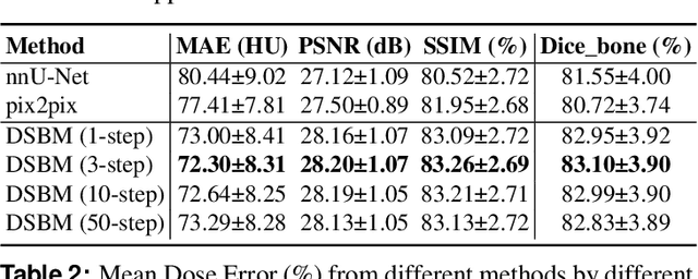
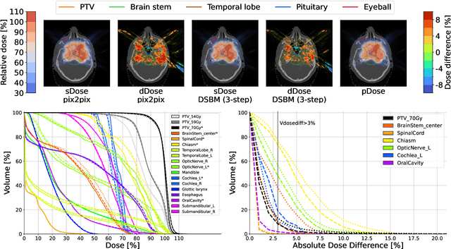
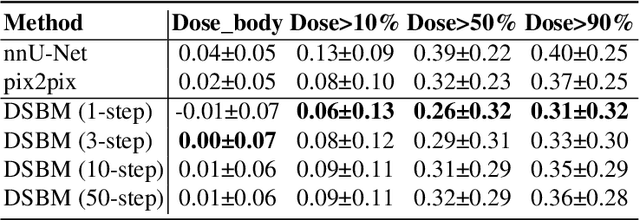
Abstract:In recent advancements in proton therapy, MR-based treatment planning is gaining momentum to minimize additional radiation exposure compared to traditional CT-based methods. This transition highlights the critical need for accurate MR-to-CT image synthesis, which is essential for precise proton dose calculations. Our research introduces the Diffusion Schr\"odinger Bridge Models (DSBM), an innovative approach for high-quality MR-to-CT synthesis. DSBM learns the nonlinear diffusion processes between MR and CT data distributions. This method improves upon traditional diffusion models by initiating synthesis from the prior distribution rather than the Gaussian distribution, enhancing both generation quality and efficiency. We validated the effectiveness of DSBM on a head and neck cancer dataset, demonstrating its superiority over traditional image synthesis methods through both image-level and dosimetric-level evaluations. The effectiveness of DSBM in MR-based proton treatment planning highlights its potential as a valuable tool in various clinical scenarios.
Neural Graphics Primitives-based Deformable Image Registration for On-the-fly Motion Extraction
Feb 08, 2024Abstract:Intra-fraction motion in radiotherapy is commonly modeled using deformable image registration (DIR). However, existing methods often struggle to balance speed and accuracy, limiting their applicability in clinical scenarios. This study introduces a novel approach that harnesses Neural Graphics Primitives (NGP) to optimize the displacement vector field (DVF). Our method leverages learned primitives, processed as splats, and interpolates within space using a shallow neural network. Uniquely, it enables self-supervised optimization at an ultra-fast speed, negating the need for pre-training on extensive datasets and allowing seamless adaptation to new cases. We validated this approach on the 4D-CT lung dataset DIR-lab, achieving a target registration error (TRE) of 1.15\pm1.15 mm within a remarkable time of 1.77 seconds. Notably, our method also addresses the sliding boundary problem, a common challenge in conventional DIR methods.
 Add to Chrome
 Add to Firefox
 Add to Edge