Alan B. McMillan
From Embeddings to Accuracy: Comparing Foundation Models for Radiographic Classification
May 16, 2025Abstract:Foundation models, pretrained on extensive datasets, have significantly advanced machine learning by providing robust and transferable embeddings applicable to various domains, including medical imaging diagnostics. This study evaluates the utility of embeddings derived from both general-purpose and medical domain-specific foundation models for training lightweight adapter models in multi-class radiography classification, focusing specifically on tube placement assessment. A dataset comprising 8842 radiographs classified into seven distinct categories was employed to extract embeddings using six foundation models: DenseNet121, BiomedCLIP, Med-Flamingo, MedImageInsight, Rad-DINO, and CXR-Foundation. Adapter models were subsequently trained using classical machine learning algorithms. Among these combinations, MedImageInsight embeddings paired with an support vector machine adapter yielded the highest mean area under the curve (mAUC) at 93.8%, followed closely by Rad-DINO (91.1%) and CXR-Foundation (89.0%). In comparison, BiomedCLIP and DenseNet121 exhibited moderate performance with mAUC scores of 83.0% and 81.8%, respectively, whereas Med-Flamingo delivered the lowest performance at 75.1%. Notably, most adapter models demonstrated computational efficiency, achieving training within one minute and inference within seconds on CPU, underscoring their practicality for clinical applications. Furthermore, fairness analyses on adapters trained on MedImageInsight-derived embeddings indicated minimal disparities, with gender differences in performance within 2% and standard deviations across age groups not exceeding 3%. These findings confirm that foundation model embeddings-especially those from MedImageInsight-facilitate accurate, computationally efficient, and equitable diagnostic classification using lightweight adapters for radiographic image analysis.
Comparative Evaluation of Radiomics and Deep Learning Models for Disease Detection in Chest Radiography
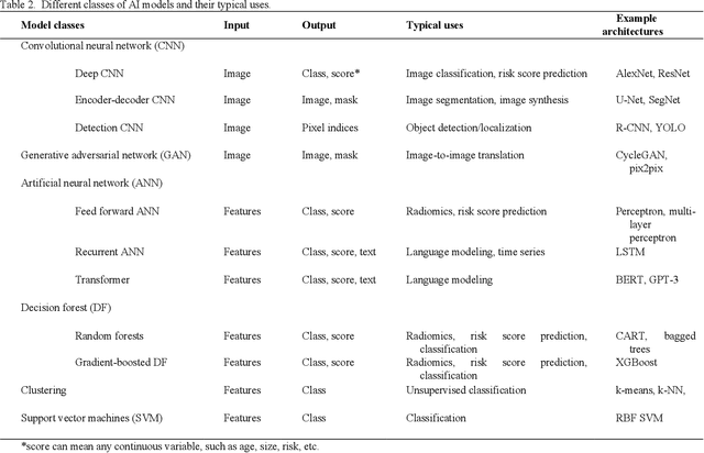
Apr 16, 2025Abstract:The application of artificial intelligence (AI) in medical imaging has revolutionized diagnostic practices, enabling advanced analysis and interpretation of radiological data. This study presents a comprehensive evaluation of radiomics-based and deep learning-based approaches for disease detection in chest radiography, focusing on COVID-19, lung opacity, and viral pneumonia. While deep learning models, particularly convolutional neural networks (CNNs) and vision transformers (ViTs), learn directly from image data, radiomics-based models extract and analyze quantitative features, potentially providing advantages in data-limited scenarios. This study systematically compares the diagnostic accuracy and robustness of various AI models, including Decision Trees, Gradient Boosting, Random Forests, Support Vector Machines (SVM), and Multi-Layer Perceptrons (MLP) for radiomics, against state-of-the-art computer vision deep learning architectures. Performance metrics across varying sample sizes reveal insights into each model's efficacy, highlighting the contexts in which specific AI approaches may offer enhanced diagnostic capabilities. The results aim to inform the integration of AI-driven diagnostic tools in clinical practice, particularly in automated and high-throughput environments where timely, reliable diagnosis is critical. This comparative study addresses an essential gap, establishing guidance for the selection of AI models based on clinical and operational needs.
Embeddings are all you need! Achieving High Performance Medical Image Classification through Training-Free Embedding Analysis
Dec 12, 2024


Abstract:Developing artificial intelligence (AI) and machine learning (ML) models for medical imaging typically involves extensive training and testing on large datasets, consuming significant computational time, energy, and resources. There is a need for more efficient methods that can achieve comparable or superior diagnostic performance without the associated resource burden. We investigated the feasibility of replacing conventional training procedures with an embedding-based approach that leverages concise and semantically meaningful representations of medical images. Using pre-trained foundational models-specifically, convolutional neural networks (CNN) like ResNet and multimodal models like Contrastive Language-Image Pre-training (CLIP)-we generated image embeddings for multi-class classification tasks. Simple linear classifiers were then applied to these embeddings. The approach was evaluated across diverse medical imaging modalities, including retinal images, mammography, dermatoscopic images, and chest radiographs. Performance was compared to benchmark models trained and tested using traditional methods. The embedding-based models surpassed the benchmark area under the receiver operating characteristic curve (AUC-ROC) scores by up to 87 percentage in multi-class classification tasks across the various medical imaging modalities. Notably, CLIP embedding models achieved the highest AUC-ROC scores, demonstrating superior classification performance while significantly reducing computational demands. Our study indicates that leveraging embeddings from pre-trained foundational models can effectively replace conventional, resource-intensive training and testing procedures in medical image analysis. This embedding-based approach offers a more efficient alternative for image segmentation, classification, and prediction, potentially accelerating AI technology integration into clinical practice.
Anatomy and Physiology of Artificial Intelligence in PET Imaging
Nov 30, 2023


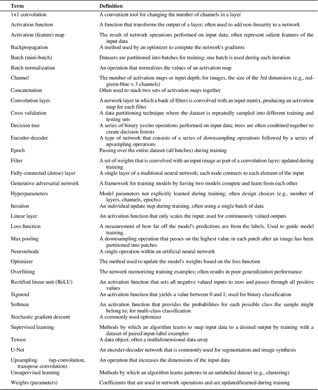
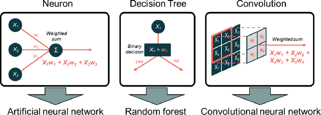
Abstract:The influence of artificial intelligence (AI) within the field of nuclear medicine has been rapidly growing. Many researchers and clinicians are seeking to apply AI within PET, and clinicians will soon find themselves engaging with AI-based applications all along the chain of molecular imaging, from image reconstruction to enhanced reporting. This expanding presence of AI in PET imaging will result in greater demand for educational resources for those unfamiliar with AI. The objective of this article to is provide an illustrated guide to the core principles of modern AI, with specific focus on aspects that are most likely to be encountered in PET imaging. We describe convolutional neural networks, algorithm training, and explain the components of the commonly used U-Net for segmentation and image synthesis.
 Add to Chrome
 Add to Firefox
 Add to Edge