 Add to Chrome
 Add to Firefox
 Add to Edge
Fundus2Globe: Generative AI-Driven 3D Digital Twins for Personalized Myopia Management
Paper and Code
Feb 18, 2025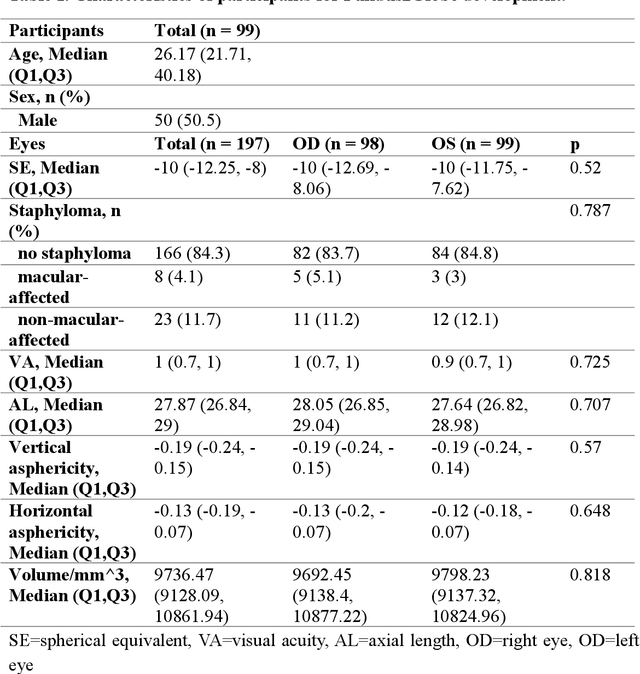
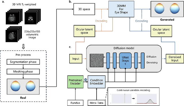
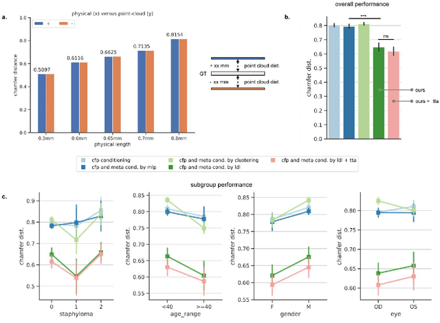
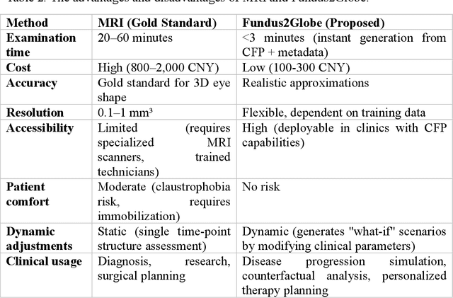
Myopia, projected to affect 50% population globally by 2050, is a leading cause of vision loss. Eyes with pathological myopia exhibit distinctive shape distributions, which are closely linked to the progression of vision-threatening complications. Recent understanding of eye-shape-based biomarkers requires magnetic resonance imaging (MRI), however, it is costly and unrealistic in routine ophthalmology clinics. We present Fundus2Globe, the first AI framework that synthesizes patient-specific 3D eye globes from ubiquitous 2D color fundus photographs (CFPs) and routine metadata (axial length, spherical equivalent), bypassing MRI dependency. By integrating a 3D morphable eye model (encoding biomechanical shape priors) with a latent diffusion model, our approach achieves submillimeter accuracy in reconstructing posterior ocular anatomy efficiently. Fundus2Globe uniquely quantifies how vision-threatening lesions (e.g., staphylomas) in CFPs correlate with MRI-validated 3D shape abnormalities, enabling clinicians to simulate posterior segment changes in response to refractive shifts. External validation demonstrates its robust generation performance, ensuring fairness across underrepresented groups. By transforming 2D fundus imaging into 3D digital replicas of ocular structures, Fundus2Globe is a gateway for precision ophthalmology, laying the foundation for AI-driven, personalized myopia management.