Ziteng Liu
C2AL: Cohort-Contrastive Auxiliary Learning for Large-scale Recommendation Systems
Oct 02, 2025


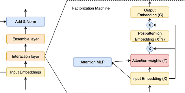
Abstract:Training large-scale recommendation models under a single global objective implicitly assumes homogeneity across user populations. However, real-world data are composites of heterogeneous cohorts with distinct conditional distributions. As models increase in scale and complexity and as more data is used for training, they become dominated by central distribution patterns, neglecting head and tail regions. This imbalance limits the model's learning ability and can result in inactive attention weights or dead neurons. In this paper, we reveal how the attention mechanism can play a key role in factorization machines for shared embedding selection, and propose to address this challenge by analyzing the substructures in the dataset and exposing those with strong distributional contrast through auxiliary learning. Unlike previous research, which heuristically applies weighted labels or multi-task heads to mitigate such biases, we leverage partially conflicting auxiliary labels to regularize the shared representation. This approach customizes the learning process of attention layers to preserve mutual information with minority cohorts while improving global performance. We evaluated C2AL on massive production datasets with billions of data points each for six SOTA models. Experiments show that the factorization machine is able to capture fine-grained user-ad interactions using the proposed method, achieving up to a 0.16% reduction in normalized entropy overall and delivering gains exceeding 0.30% on targeted minority cohorts.
Monocular Depth Guided Occlusion-Aware Disparity Refinement via Semi-supervised Learning in Laparoscopic Images
May 13, 2025Abstract:Occlusion and the scarcity of labeled surgical data are significant challenges in disparity estimation for stereo laparoscopic images. To address these issues, this study proposes a Depth Guided Occlusion-Aware Disparity Refinement Network (DGORNet), which refines disparity maps by leveraging monocular depth information unaffected by occlusion. A Position Embedding (PE) module is introduced to provide explicit spatial context, enhancing the network's ability to localize and refine features. Furthermore, we introduce an Optical Flow Difference Loss (OFDLoss) for unlabeled data, leveraging temporal continuity across video frames to improve robustness in dynamic surgical scenes. Experiments on the SCARED dataset demonstrate that DGORNet outperforms state-of-the-art methods in terms of End-Point Error (EPE) and Root Mean Squared Error (RMSE), particularly in occlusion and texture-less regions. Ablation studies confirm the contributions of the Position Embedding and Optical Flow Difference Loss, highlighting their roles in improving spatial and temporal consistency. These results underscore DGORNet's effectiveness in enhancing disparity estimation for laparoscopic surgery, offering a practical solution to challenges in disparity estimation and data limitations.
Attention-Aware Laparoscopic Image Desmoking Network with Lightness Embedding and Hybrid Guided Embedding
Apr 11, 2024


Abstract:This paper presents a novel method of smoke removal from the laparoscopic images. Due to the heterogeneous nature of surgical smoke, a two-stage network is proposed to estimate the smoke distribution and reconstruct a clear, smoke-free surgical scene. The utilization of the lightness channel plays a pivotal role in providing vital information pertaining to smoke density. The reconstruction of smoke-free image is guided by a hybrid embedding, which combines the estimated smoke mask with the initial image. Experimental results demonstrate that the proposed method boasts a Peak Signal to Noise Ratio that is $2.79\%$ higher than the state-of-the-art methods, while also exhibits a remarkable $38.2\%$ reduction in run-time. Overall, the proposed method offers comparable or even superior performance in terms of both smoke removal quality and computational efficiency when compared to existing state-of-the-art methods. This work will be publicly available on http://homepage.hit.edu.cn/wpgao
Self-Supervised Surgical Instrument 3D Reconstruction from a Single Camera Image
Nov 26, 2022Abstract:Surgical instrument tracking is an active research area that can provide surgeons feedback about the location of their tools relative to anatomy. Recent tracking methods are mainly divided into two parts: segmentation and object detection. However, both can only predict 2D information, which is limiting for application to real-world surgery. An accurate 3D surgical instrument model is a prerequisite for precise predictions of the pose and depth of the instrument. Recent single-view 3D reconstruction methods are only used in natural object reconstruction and do not achieve satisfying reconstruction accuracy without 3D attribute-level supervision. Further, those methods are not suitable for the surgical instruments because of their elongated shapes. In this paper, we firstly propose an end-to-end surgical instrument reconstruction system -- Self-supervised Surgical Instrument Reconstruction (SSIR). With SSIR, we propose a multi-cycle-consistency strategy to help capture the texture information from a slim instrument while only requiring a binary instrument label map. Experiments demonstrate that our approach improves the reconstruction quality of surgical instruments compared to other self-supervised methods and achieves promising results.
Min-Max Similarity: A Contrastive Learning Based Semi-Supervised Learning Network for Surgical Tools Segmentation
Mar 29, 2022


Abstract:Segmentation of images is a popular topic in medical AI. This is mainly due to the difficulty to obtain a significant number of pixel-level annotated data to train a neural network. To address this issue, we proposed a semi-supervised segmentation network based on contrastive learning. In contrast to the previous state-of-the-art, we introduce a contrastive learning form of dual-view training by employing classifiers and projectors to build all-negative, and positive and negative feature pairs respectively to formulate the learning problem as solving min-max similarity problem. The all-negative pairs are used to supervise the networks learning from different views and make sure to capture general features, and the consistency of unlabeled predictions is measured by pixel-wise contrastive loss between positive and negative pairs. To quantitative and qualitative evaluate our proposed method, we test it on two public endoscopy surgical tool segmentation datasets and one cochlear implant surgery dataset which we manually annotate the cochlear implant in surgical videos. The segmentation performance (dice coefficients) indicates that our proposed method outperforms state-of-the-art semi-supervised and fully supervised segmentation algorithms consistently. The code is publicly available at: https://github.com/AngeLouCN/Min_Max_Similarity
 Add to Chrome
 Add to Firefox
 Add to Edge