Zahra Sobhaninia
Endoscopy Classification Model Using Swin Transformer and Saliency Map
Mar 12, 2023


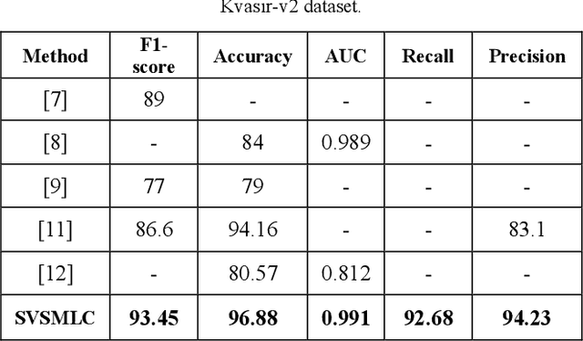
Abstract:Endoscopy is a valuable tool for the early diagnosis of colon cancer. However, it requires the expertise of endoscopists and is a time-consuming process. In this work, we propose a new multi-label classification method, which considers two aspects of learning approaches (local and global views) for endoscopic image classification. The model consists of a Swin transformer branch and a modified VGG16 model as a CNN branch. To help the learning process of the CNN branch, the model employs saliency maps and endoscopy images and concatenates them. The results demonstrate that this method performed well for endoscopic medical images by utilizing local and global features of the images. Furthermore, quantitative evaluations prove the proposed method's superiority over state-of-the-art works.
Brain Tumor Classification by Cascaded Multiscale Multitask Learning Framework Based on Feature Aggregation
Dec 28, 2021


Abstract:Brain tumor analysis in MRI images is a significant and challenging issue because misdiagnosis can lead to death. Diagnosis and evaluation of brain tumors in the early stages increase the probability of successful treatment. However, the complexity and variety of tumors, shapes, and locations make their segmentation and classification complex. In this regard, numerous researchers have proposed brain tumor segmentation and classification methods. This paper presents an approach that simultaneously segments and classifies brain tumors in MRI images using a framework that contains MRI image enhancement and tumor region detection. Eventually, a network based on a multitask learning approach is proposed. Subjective and objective results indicate that the segmentation and classification results based on evaluation metrics are better or comparable to the state-of-the-art.
Brain Tumor Segmentation by Cascaded Deep Neural Networks Using Multiple Image Scales
Feb 05, 2020
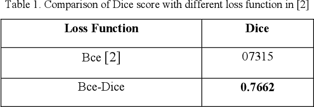

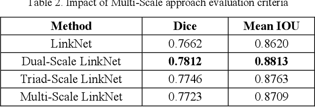
Abstract:Intracranial tumors are groups of cells that usually grow uncontrollably. One out of four cancer deaths is due to brain tumors. Early detection and evaluation of brain tumors is an essential preventive medical step that is performed by magnetic resonance imaging (MRI). Many segmentation techniques exist for this purpose. Low segmentation accuracy is the main drawback of existing methods. In this paper, we use a deep learning method to boost the accuracy of tumor segmentation in MR images. Cascade approach is used with multiple scales of images to induce both local and global views and help the network to reach higher accuracies. Our experimental results show that using multiple scales and the utilization of two cascade networks is advantageous.
Localization of Fetal Head in Ultrasound Images by Multiscale View and Deep Neural Networks
Nov 03, 2019


Abstract:One of the routine examinations that are used for prenatal care in many countries is ultrasound imaging. This procedure provides various information about fetus health and development, the progress of the pregnancy and, the baby's due date. Some of the biometric parameters of the fetus, like fetal head circumference (HC), must be measured to check the fetus's health and growth. In this paper, we investigated the effects of using multi-scale inputs in the network. We also propose a light convolutional neural network for automatic HC measurement. Experimental results on an ultrasound dataset of the fetus in different trimesters of pregnancy show that the segmentation accuracy and HC evaluations performed by a light convolutional neural network are comparable to deep convolutional neural networks. The proposed network has fewer parameters and requires less training time.
Fetal Ultrasound Image Segmentation for Measuring Biometric Parameters Using Multi-Task Deep Learning
Aug 31, 2019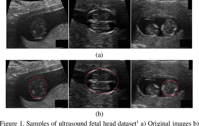
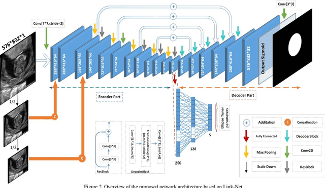
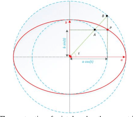

Abstract:Ultrasound imaging is a standard examination during pregnancy that can be used for measuring specific biometric parameters towards prenatal diagnosis and estimating gestational age. Fetal head circumference (HC) is one of the significant factors to determine the fetus growth and health. In this paper, a multi-task deep convolutional neural network is proposed for automatic segmentation and estimation of HC ellipse by minimizing a compound cost function composed of segmentation dice score and MSE of ellipse parameters. Experimental results on fetus ultrasound dataset in different trimesters of pregnancy show that the segmentation results and the extracted HC match well with the radiologist annotations. The obtained dice scores of the fetal head segmentation and the accuracy of HC evaluations are comparable to the state-of-the-art.
Brain Tumor Segmentation Using Deep Learning by Type Specific Sorting of Images
Sep 20, 2018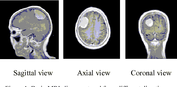
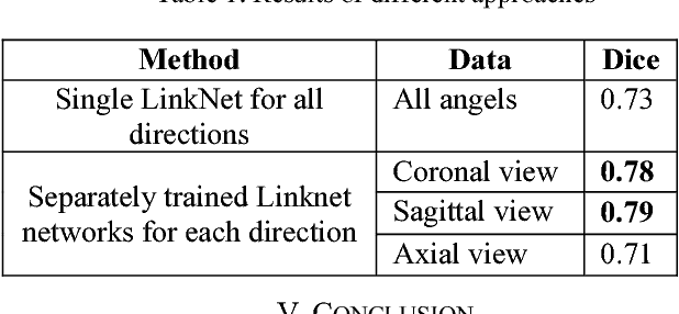

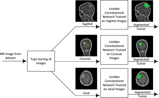
Abstract:Recently deep learning has been playing a major role in the field of computer vision. One of its applications is the reduction of human judgment in the diagnosis of diseases. Especially, brain tumor diagnosis requires high accuracy, where minute errors in judgment may lead to disaster. For this reason, brain tumor segmentation is an important challenge for medical purposes. Currently several methods exist for tumor segmentation but they all lack high accuracy. Here we present a solution for brain tumor segmenting by using deep learning. In this work, we studied different angles of brain MR images and applied different networks for segmentation. The effect of using separate networks for segmentation of MR images is evaluated by comparing the results with a single network. Experimental evaluations of the networks show that Dice score of 0.73 is achieved for a single network and 0.79 in obtained for multiple networks.
 Add to Chrome
 Add to Firefox
 Add to Edge