Yernar Zhetpissov
A-SEE2.0: Active-Sensing End-Effector for Robotic Ultrasound Systems with Dense Contact Surface Perception Enabled Probe Orientation Adjustment
Mar 07, 2025


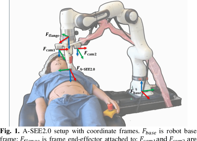
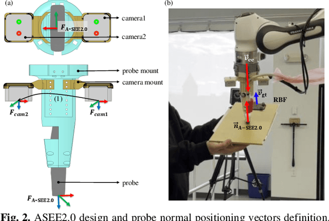
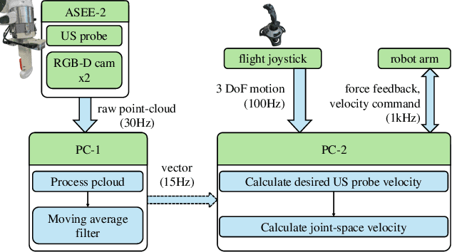
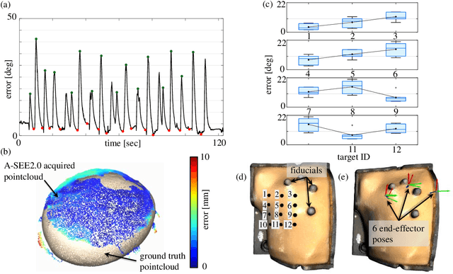
Abstract:Conventional freehand ultrasound (US) imaging is highly dependent on the skill of the operator, often leading to inconsistent results and increased physical demand on sonographers. Robotic Ultrasound Systems (RUSS) aim to address these limitations by providing standardized and automated imaging solutions, especially in environments with limited access to skilled operators. This paper presents the development of a novel RUSS system that employs dual RGB-D depth cameras to maintain the US probe normal to the skin surface, a critical factor for optimal image quality. Our RUSS integrates RGB-D camera data with robotic control algorithms to maintain orthogonal probe alignment on uneven surfaces without preoperative data. Validation tests using a phantom model demonstrate that the system achieves robust normal positioning accuracy while delivering ultrasound images comparable to those obtained through manual scanning. A-SEE2.0 demonstrates 2.47 ${\pm}$ 1.25 degrees error for flat surface normal-positioning and 12.19 ${\pm}$ 5.81 degrees normal estimation error on mannequin surface. This work highlights the potential of A-SEE2.0 to be used in clinical practice by testing its performance during in-vivo forearm ultrasound examinations.
Optical Fiber-Based Needle Shape Sensing in Real Tissue: Single Core vs. Multicore Approaches
Sep 08, 2023


Abstract:Flexible needle insertion procedures are common for minimally-invasive surgeries for diagnosing and treating prostate cancer. Bevel-tip needles provide physicians the capability to steer the needle during long insertions to avoid vital anatomical structures in the patient and reduce post-operative patient discomfort. To provide needle placement feedback to the physician, sensors are embedded into needles for determining the real-time 3D shape of the needle during operation without needing to visualize the needle intra-operatively. Through expansive research in fiber optics, a plethora of bio-compatible, MRI-compatible, optical shape-sensors have been developed to provide real-time shape feedback, such as single-core and multicore fiber Bragg gratings. In this paper, we directly compare single-core fiber-based and multicore fiber-based needle shape-sensing through identically constructed, four-active area sensorized bevel-tip needles inserted into phantom and \exvivo tissue on the same experimental platform. In this work, we found that for shape-sensing in phantom tissue, the two needles performed identically with a $p$-value of $0.164 > 0.05$, but in \exvivo real tissue, the single-core fiber sensorized needle significantly outperformed the multicore fiber configuration with a $p$-value of $0.0005 < 0.05$. This paper also presents the experimental platform and method for directly comparing these optical shape sensors for the needle shape-sensing task, as well as provides direction, insight and required considerations for future work in constructively optimizing sensorized needles.
 Add to Chrome
 Add to Firefox
 Add to Edge