Xiaohong Ma
Deep Distance Map Regression Network with Shape-aware Loss for Imbalanced Medical Image Segmentation
Jan 15, 2025Abstract:Small object segmentation, like tumor segmentation, is a difficult and critical task in the field of medical image analysis. Although deep learning based methods have achieved promising performance, they are restricted to the use of binary segmentation mask. Inspired by the rigorous mapping between binary segmentation mask and distance map, we adopt distance map as a novel ground truth and employ a network to fulfill the computation of distance map. Specially, we propose a new segmentation framework that incorporates the existing binary segmentation network and a light weight regression network (dubbed as LR-Net). Thus, the LR-Net can convert the distance map computation into a regression task and leverage the rich information of distance maps. Additionally, we derive a shape-aware loss by employing distance maps as penalty map to infer the complete shape of an object. We evaluated our approach on MICCAI 2017 Liver Tumor Segmentation (LiTS) Challenge dataset and a clinical dataset. Experimental results show that our approach outperforms the classification-based methods as well as other existing state-of-the-arts.
* Conference
Fusing Medical Image Features and Clinical Features with Deep Learning for Computer-Aided Diagnosis
Mar 10, 2021


Abstract:Current Computer-Aided Diagnosis (CAD) methods mainly depend on medical images. The clinical information, which usually needs to be considered in practical clinical diagnosis, has not been fully employed in CAD. In this paper, we propose a novel deep learning-based method for fusing Magnetic Resonance Imaging (MRI)/Computed Tomography (CT) images and clinical information for diagnostic tasks. Two paths of neural layers are performed to extract image features and clinical features, respectively, and at the same time clinical features are employed as the attention to guide the extraction of image features. Finally, these two modalities of features are concatenated to make decisions. We evaluate the proposed method on its applications to Alzheimer's disease diagnosis, mild cognitive impairment converter prediction and hepatic microvascular invasion diagnosis. The encouraging experimental results prove the values of the image feature extraction guided by clinical features and the concatenation of two modalities of features for classification, which improve the performance of diagnosis effectively and stably.
A New Computer-Aided Diagnosis System with Modified Genetic Feature Selection for BI-RADS Classification of Breast Masses in Mammograms
May 11, 2020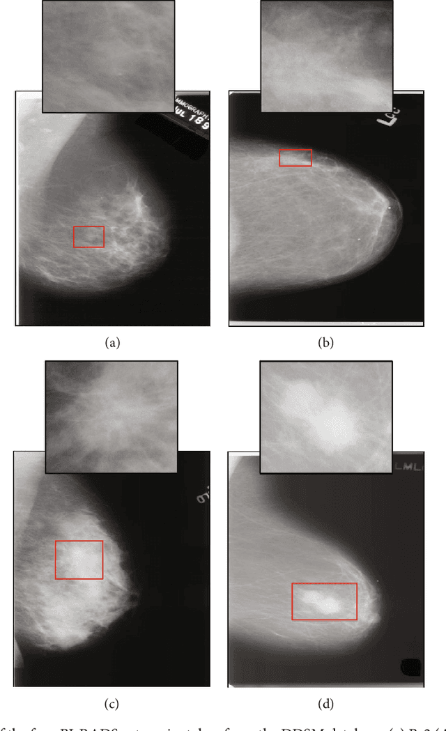
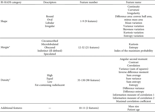

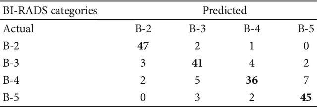
Abstract:Mammography remains the most prevalent imaging tool for early breast cancer screening. The language used to describe abnormalities in mammographic reports is based on the breast Imaging Reporting and Data System (BI-RADS). Assigning a correct BI-RADS category to each examined mammogram is a strenuous and challenging task for even experts. This paper proposes a new and effective computer-aided diagnosis (CAD) system to classify mammographic masses into four assessment categories in BI-RADS. The mass regions are first enhanced by means of histogram equalization and then semiautomatically segmented based on the region growing technique. A total of 130 handcrafted BI-RADS features are then extrcated from the shape, margin, and density of each mass, together with the mass size and the patient's age, as mentioned in BI-RADS mammography. Then, a modified feature selection method based on the genetic algorithm (GA) is proposed to select the most clinically significant BI-RADS features. Finally, a back-propagation neural network (BPN) is employed for classification, and its accuracy is used as the fitness in GA. A set of 500 mammogram images from the digital database of screening mammography (DDSM) is used for evaluation. Our system achieves classification accuracy, positive predictive value, negative predictive value, and Matthews correlation coefficient of 84.5%, 84.4%, 94.8%, and 79.3%, respectively. To our best knowledge, this is the best current result for BI-RADS classification of breast masses in mammography, which makes the proposed system promising to support radiologists for deciding proper patient management based on the automatically assigned BI-RADS categories.
A New Three-stage Curriculum Learning Approach to Deep Network Based Liver Tumor Segmentation
Oct 17, 2019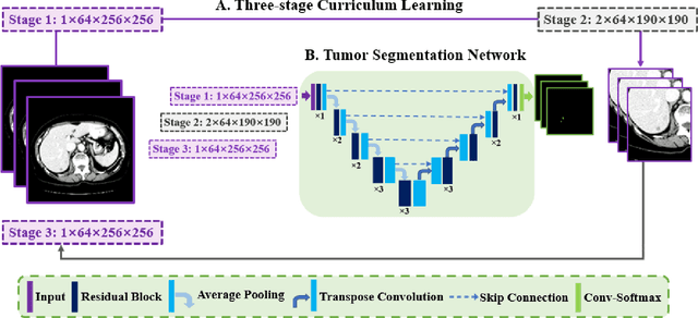

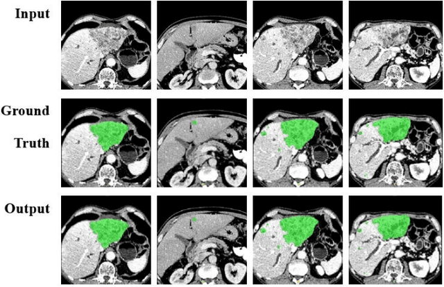
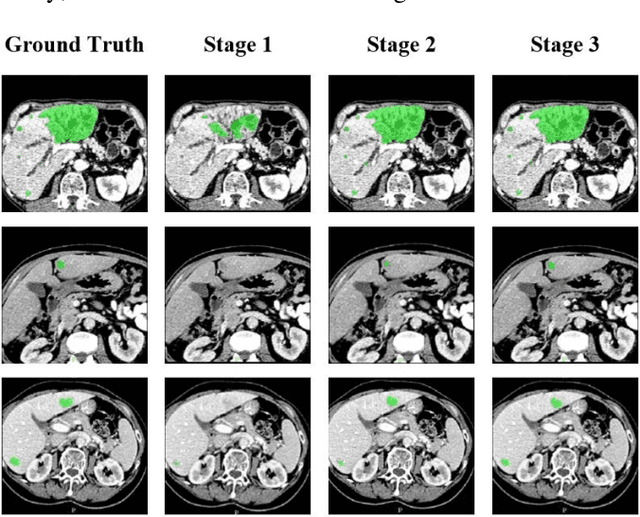
Abstract:Automatic segmentation of liver tumors in medical images is crucial for the computer-aided diagnosis and therapy. It is a challenging task, since the tumors are notoriously small against the background voxels. This paper proposes a new three-stage curriculum learning approach for training deep networks to tackle this small object segmentation problem. The learning in the first stage is performed on the whole input to obtain an initial deep network for tumor segmenta-tion. Then the second stage of learning focuses the strength-ening of tumor specific features by continuing training the network on the tumor patches. Finally, we retrain the net-work on the whole input in the third stage, in order that the tumor specific features and the global context can be inte-grated ideally under the segmentation objective. Benefitting from the proposed learning approach, we only need to em-ploy one single network to segment the tumors directly. We evaluated our approach on the 2017 MICCAI Liver Tumor Segmentation challenge dataset. In the experiments, our approach exhibits significant improvement compared with the commonly used cascaded counterpart.
 Add to Chrome
 Add to Firefox
 Add to Edge