Tanmoy Debnath
A Sparse-Attention Deep Learning Model Integrating Heterogeneous Multimodal Features for Parkinson's Disease Severity Profiling
Jan 02, 2026Abstract:Characterising the heterogeneous presentation of Parkinson's disease (PD) requires integrating biological and clinical markers within a unified predictive framework. While multimodal data provide complementary information, many existing computational models struggle with interpretability, class imbalance, or effective fusion of high-dimensional imaging and tabular clinical features. To address these limitations, we propose the Class-Weighted Sparse-Attention Fusion Network (SAFN), an interpretable deep learning framework for robust multimodal profiling. SAFN integrates MRI cortical thickness, MRI volumetric measures, clinical assessments, and demographic variables using modality-specific encoders and a symmetric cross-attention mechanism that captures nonlinear interactions between imaging and clinical representations. A sparsity-constrained attention-gating fusion layer dynamically prioritises informative modalities, while a class-balanced focal loss (beta = 0.999, gamma = 1.5) mitigates dataset imbalance without synthetic oversampling. Evaluated on 703 participants (570 PD, 133 healthy controls) from the Parkinson's Progression Markers Initiative using subject-wise five-fold cross-validation, SAFN achieves an accuracy of 0.98 plus or minus 0.02 and a PR-AUC of 1.00 plus or minus 0.00, outperforming established machine learning and deep learning baselines. Interpretability analysis shows a clinically coherent decision process, with approximately 60 percent of predictive weight assigned to clinical assessments, consistent with Movement Disorder Society diagnostic principles. SAFN provides a reproducible and transparent multimodal modelling paradigm for computational profiling of neurodegenerative disease.
Efficient quantum image representation and compression circuit using zero-discarded state preparation approach
Jun 22, 2023


Abstract:Quantum image computing draws a lot of attention due to storing and processing image data faster than classical. With increasing the image size, the number of connections also increases, leading to the circuit complex. Therefore, efficient quantum image representation and compression issues are still challenging. The encoding of images for representation and compression in quantum systems is different from classical ones. In quantum, encoding of position is more concerned which is the major difference from the classical. In this paper, a novel zero-discarded state connection novel enhance quantum representation (ZSCNEQR) approach is introduced to reduce complexity further by discarding '0' in the location representation information. In the control operational gate, only input '1' contribute to its output thus, discarding zero makes the proposed ZSCNEQR circuit more efficient. The proposed ZSCNEQR approach significantly reduced the required bit for both representation and compression. The proposed method requires 11.76\% less qubits compared to the recent existing method. The results show that the proposed approach is highly effective for representing and compressing images compared to the two relevant existing methods in terms of rate-distortion performance.
A novel state connection strategy for quantum computing to represent and compress digital images
Dec 14, 2022


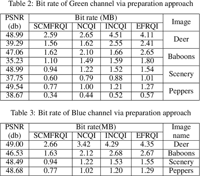
Abstract:Quantum image processing draws a lot of attention due to faster data computation and storage compared to classical data processing systems. Converting classical image data into the quantum domain and state label preparation complexity is still a challenging issue. The existing techniques normally connect the pixel values and the state position directly. Recently, the EFRQI (efficient flexible representation of the quantum image) approach uses an auxiliary qubit that connects the pixel-representing qubits to the state position qubits via Toffoli gates to reduce state connection. Due to the twice use of Toffoli gates for each pixel connection still it requires a significant number of bits to connect each pixel value. In this paper, we propose a new SCMFRQI (state connection modification FRQI) approach for further reducing the required bits by modifying the state connection using a reset gate rather than repeating the use of the same Toffoli gate connection as a reset gate. Moreover, unlike other existing methods, we compress images using block-level for further reduction of required qubits. The experimental results confirm that the proposed method outperforms the existing methods in terms of both image representation and compression points of view.
Advance quantum image representation and compression using DCTEFRQI approach
Aug 30, 2022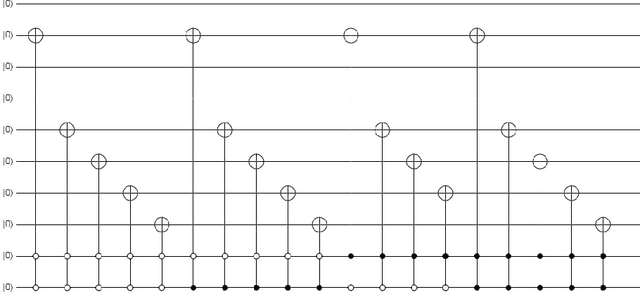
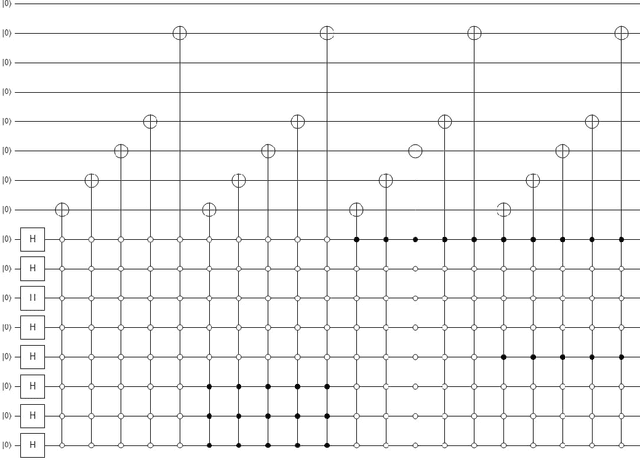
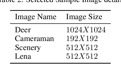
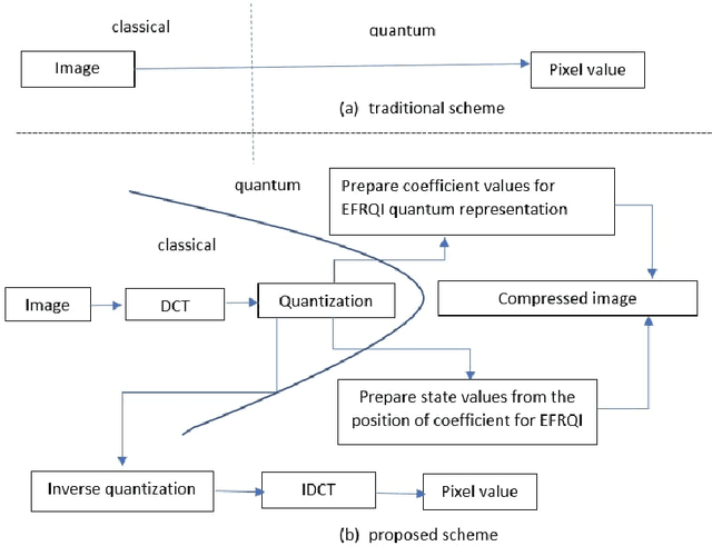
Abstract:In recent year, quantum image processing got a lot of attention in the field of image processing due to opportunity to place huge image data in quantum Hilbert space. Hilbert space or Euclidean space has infinite dimension to locate and process the image data faster. Moreover, several researches show that, the computational time of quantum process is faster than classical computer. By encoding and compressing the image in quantum domain is still challenging issue. From literature survey, we have proposed a DCTEFRQI (Direct Cosine Transform Efficient Flexible Representation of Quantum Image) algorithm to represent and compress gray image efficiently which save computational time and minimize the complexity of preparation. The objective of this work is to represent and compress various gray image size in quantum computer using DCT(Discrete Cosine Transform) and EFRQI (Efficient Flexible Representation of Quantum Image) approach together. Quirk simulation tool is used to design corresponding quantum image circuit. Due to limitation of qubit, total 16 numbers of qubit are used to represent the gray scale image among those 8 are used to map the coefficient values and the rest 8 are used to generate the corresponding coefficient position. Theoretical analysis and experimental result show that, proposed DCTEFRQI scheme provides better representation and compression compare to DCT-GQIR, DWT-GQIR and DWT-EFRQI in terms of PSNR(Peak Signal to Noise Ratio) and bit rate..
MAVIDH Score: A COVID-19 Severity Scoring using Chest X-Ray Pathology Features
Dec 01, 2020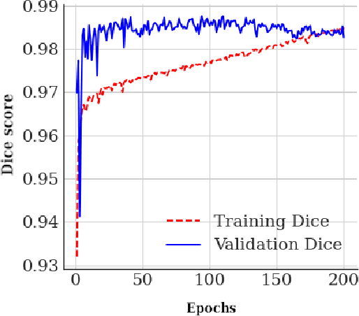
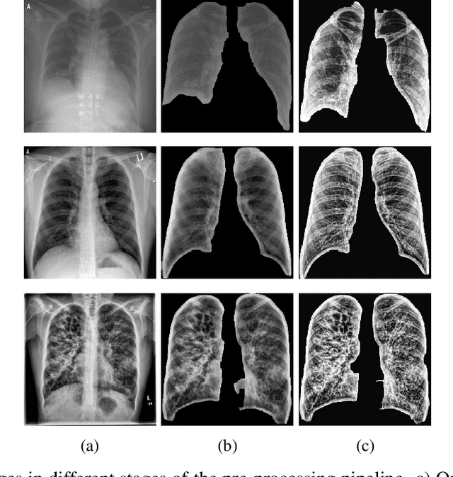
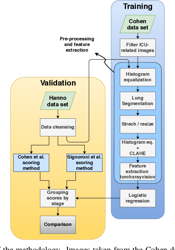
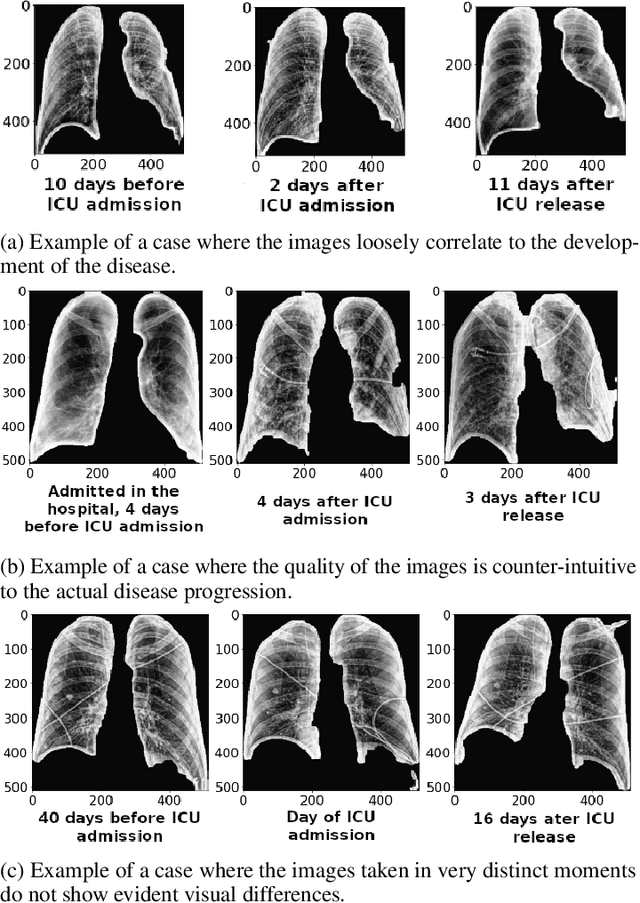
Abstract:The application of computer vision for COVID-19 diagnosis is complex and challenging, given the risks associated with patient misclassifications. Arguably, the primary value of medical imaging for COVID-19 lies rather on patient prognosis. Radiological images can guide physicians assessing the severity of the disease, and a series of images from the same patient at different stages can help to gauge disease progression. Based on these premises, a simple method based on lung-pathology features for scoring disease severity from Chest X-rays is proposed here. As the primary contribution, this method shows to be correlated to patient severity in different stages of disease progression comparatively well when contrasted with other existing methods. An original approach for data selection is also proposed, allowing the simple model to learn the severity-related features. It is hypothesized that the resulting competitive performance presented here is related to the method being feature-based rather than reliant on lung involvement or compromise as others in the literature. The fact that it is simpler and interpretable than other end-to-end, more complex models, also sets aside this work. As the data set is small, bias-inducing artifacts that could lead to overfitting are minimized through an image normalization and lung segmentation step at the learning phase. A second contribution comes from the validation of the results, conceptualized as the scoring of patients groups from different stages of the disease. Besides performing such validation on an independent data set, the results were also compared with other proposed scoring methods in the literature. The expressive results show that although imaging alone is not sufficient for assessing severity as a whole, there is a strong correlation with the scoring system, termed as MAVIDH score, with patient outcome.
Potential Features of ICU Admission in X-ray Images of COVID-19 Patients
Sep 26, 2020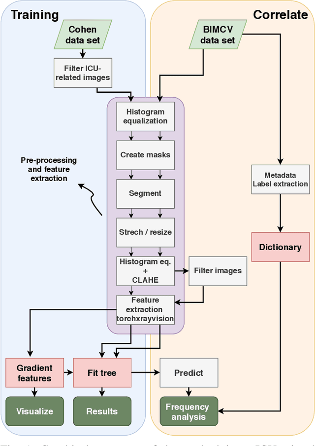
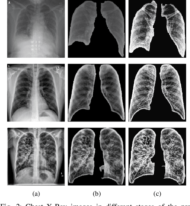
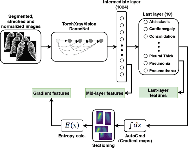
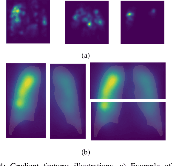
Abstract:X-ray images may present non-trivial features with predictive information of patients that develop severe symptoms of COVID-19. If true, this hypothesis may have practical value in allocating resources to particular patients while using a relatively inexpensive imaging technique. The difficulty of testing such a hypothesis comes from the need for large sets of labelled data, which not only need to be well-annotated but also should contemplate the post-imaging severity outcome. On this account, this paper presents a methodology for extracting features from a limited data set with outcome label (patient required ICU admission or not) and correlating its significance to an additional, larger data set with hundreds of images. The methodology employs a neural network trained to recognise lung pathologies to extract the semantic features, which are then analysed with a shallow decision tree to limit overfitting while increasing interpretability. This analysis points out that only a few features explain most of the variance between patients that developed severe symptoms. When applied to an unrelated, larger data set with labels extracted from clinical notes, the method classified distinct sets of samples where there was a much higher frequency of labels such as `Consolidation', `Effusion', and `alveolar'. A further brief analysis on the locations of such labels also showed a significant increase in the frequency of words like `bilateral', `middle', and `lower' in patients classified as with higher chances of going severe. The methodology for dealing with the lack of specific ICU label data while attesting correlations with a data set containing text notes is novel; its results suggest that some pathologies should receive higher weights when assessing disease severity.
Hyperspectral Imaging to detect Age, Defects and Individual Nutrient Deficiency in Grapevine Leaves
Jul 10, 2020
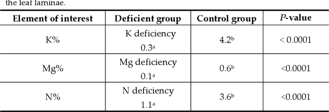


Abstract:Hyperspectral (HS) imaging was successfully employed in the 380 nm to 1000 nm wavelength range to investigate the efficacy of detecting age, healthiness and individual nutrient deficiency of grapevine leaves collected from vineyards located in central west NSW, Australia. For age detection, the appearance of many healthy grapevine leaves has been examined. Then visually defective leaves were compared with healthy leaves. Control leaves and individual nutrient-deficient leaves (e.g. N, K and Mg) were also analysed. Several features were employed at various stages in the Ultraviolet (UV), Visible (VIS) and Near Infrared (NIR) regions to evaluate the experimental data: mean brightness, mean 1st derivative brightness, variation index, mean spectral ratio, normalised difference vegetation index (NDVI) and standard deviation (SD). Experiment results demonstrate that these features could be utilised with a high degree of effectiveness to compare age, identify unhealthy samples and not only to distinguish from control and nutrient deficiency but also to identify individual nutrient defects. Therefore, our work corroborated that HS imaging has excellent potential as a non-destructive as well as a non-contact method to detect age, healthiness and individual nutrient deficiencies of grapevine leaves
 Add to Chrome
 Add to Firefox
 Add to Edge