Tabea-Clara Bucher
Pathologist-like explainable AI for interpretable Gleason grading in prostate cancer
Oct 19, 2024


Abstract:The aggressiveness of prostate cancer, the most common cancer in men worldwide, is primarily assessed based on histopathological data using the Gleason scoring system. While artificial intelligence (AI) has shown promise in accurately predicting Gleason scores, these predictions often lack inherent explainability, potentially leading to distrust in human-machine interactions. To address this issue, we introduce a novel dataset of 1,015 tissue microarray core images, annotated by an international group of 54 pathologists. The annotations provide detailed localized pattern descriptions for Gleason grading in line with international guidelines. Utilizing this dataset, we develop an inherently explainable AI system based on a U-Net architecture that provides predictions leveraging pathologists' terminology. This approach circumvents post-hoc explainability methods while maintaining or exceeding the performance of methods trained directly for Gleason pattern segmentation (Dice score: 0.713 $\pm$ 0.003 trained on explanations vs. 0.691 $\pm$ 0.010 trained on Gleason patterns). By employing soft labels during training, we capture the intrinsic uncertainty in the data, yielding strong results in Gleason pattern segmentation even in the context of high interobserver variability. With the release of this dataset, we aim to encourage further research into segmentation in medical tasks with high levels of subjectivity and to advance the understanding of pathologists' reasoning processes.
On the calibration of neural networks for histological slide-level classification
Dec 15, 2023
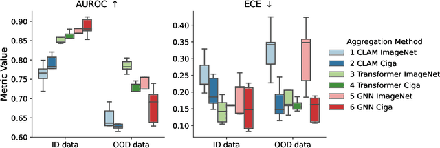
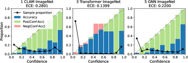

Abstract:Deep Neural Networks have shown promising classification performance when predicting certain biomarkers from Whole Slide Images in digital pathology. However, the calibration of the networks' output probabilities is often not evaluated. Communicating uncertainty by providing reliable confidence scores is of high relevance in the medical context. In this work, we compare three neural network architectures that combine feature representations on patch-level to a slide-level prediction with respect to their classification performance and evaluate their calibration. As slide-level classification task, we choose the prediction of Microsatellite Instability from Colorectal Cancer tissue sections. We observe that Transformers lead to good results in terms of classification performance and calibration. When evaluating the classification performance on a separate dataset, we observe that Transformers generalize best. The investigation of reliability diagrams provides additional insights to the Expected Calibration Error metric and we observe that especially Transformers push the output probabilities to extreme values, which results in overconfident predictions.
Evaluating Deep Learning-based Melanoma Classification using Immunohistochemistry and Routine Histology: A Three Center Study
Sep 08, 2023


Abstract:Pathologists routinely use immunohistochemical (IHC)-stained tissue slides against MelanA in addition to hematoxylin and eosin (H&E)-stained slides to improve their accuracy in diagnosing melanomas. The use of diagnostic Deep Learning (DL)-based support systems for automated examination of tissue morphology and cellular composition has been well studied in standard H&E-stained tissue slides. In contrast, there are few studies that analyze IHC slides using DL. Therefore, we investigated the separate and joint performance of ResNets trained on MelanA and corresponding H&E-stained slides. The MelanA classifier achieved an area under receiver operating characteristics curve (AUROC) of 0.82 and 0.74 on out of distribution (OOD)-datasets, similar to the H&E-based benchmark classification of 0.81 and 0.75, respectively. A combined classifier using MelanA and H&E achieved AUROCs of 0.85 and 0.81 on the OOD datasets. DL MelanA-based assistance systems show the same performance as the benchmark H&E classification and may be improved by multi stain classification to assist pathologists in their clinical routine.
Dermatologist-like explainable AI enhances trust and confidence in diagnosing melanoma
Mar 17, 2023Abstract:Although artificial intelligence (AI) systems have been shown to improve the accuracy of initial melanoma diagnosis, the lack of transparency in how these systems identify melanoma poses severe obstacles to user acceptance. Explainable artificial intelligence (XAI) methods can help to increase transparency, but most XAI methods are unable to produce precisely located domain-specific explanations, making the explanations difficult to interpret. Moreover, the impact of XAI methods on dermatologists has not yet been evaluated. Extending on two existing classifiers, we developed an XAI system that produces text and region based explanations that are easily interpretable by dermatologists alongside its differential diagnoses of melanomas and nevi. To evaluate this system, we conducted a three-part reader study to assess its impact on clinicians' diagnostic accuracy, confidence, and trust in the XAI-support. We showed that our XAI's explanations were highly aligned with clinicians' explanations and that both the clinicians' trust in the support system and their confidence in their diagnoses were significantly increased when using our XAI compared to using a conventional AI system. The clinicians' diagnostic accuracy was numerically, albeit not significantly, increased. This work demonstrates that clinicians are willing to adopt such an XAI system, motivating their future use in the clinic.
Multi-domain stain normalization for digital pathology: A cycle-consistent adversarial network for whole slide images
Jan 23, 2023


Abstract:The variation in histologic staining between different medical centers is one of the most profound challenges in the field of computer-aided diagnosis. The appearance disparity of pathological whole slide images causes algorithms to become less reliable, which in turn impedes the wide-spread applicability of downstream tasks like cancer diagnosis. Furthermore, different stainings lead to biases in the training which in case of domain shifts negatively affect the test performance. Therefore, in this paper we propose MultiStain-CycleGAN, a multi-domain approach to stain normalization based on CycleGAN. Our modifications to CycleGAN allow us to normalize images of different origins without retraining or using different models. We perform an extensive evaluation of our method using various metrics and compare it to commonly used methods that are multi-domain capable. First, we evaluate how well our method fools a domain classifier that tries to assign a medical center to an image. Then, we test our normalization on the tumor classification performance of a downstream classifier. Furthermore, we evaluate the image quality of the normalized images using the Structural similarity index and the ability to reduce the domain shift using the Fr\'echet inception distance. We show that our method proves to be multi-domain capable, provides the highest image quality among the compared methods, and can most reliably fool the domain classifier while keeping the tumor classifier performance high. By reducing the domain influence, biases in the data can be removed on the one hand and the origin of the whole slide image can be disguised on the other, thus enhancing patient data privacy.
Benchmarking common uncertainty estimation methods with histopathological images under domain shift and label noise
Jan 03, 2023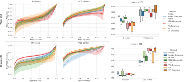
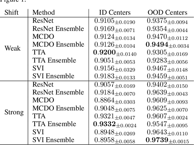

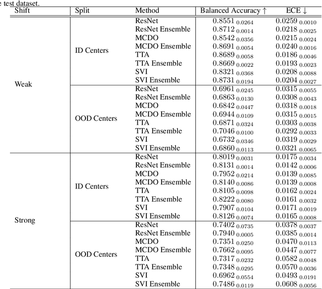
Abstract:In the past years, deep learning has seen an increase of usage in the domain of histopathological applications. However, while these approaches have shown great potential, in high-risk environments deep learning models need to be able to judge their own uncertainty and be able to reject inputs when there is a significant chance of misclassification. In this work, we conduct a rigorous evaluation of the most commonly used uncertainty and robustness methods for the classification of Whole-Slide-Images under domain shift using the H\&E stained Camelyon17 breast cancer dataset. Although it is known that histopathological data can be subject to strong domain shift and label noise, to our knowledge this is the first work that compares the most common methods for uncertainty estimation under these aspects. In our experiments, we compare Stochastic Variational Inference, Monte-Carlo Dropout, Deep Ensembles, Test-Time Data Augmentation as well as combinations thereof. We observe that ensembles of methods generally lead to higher accuracies and better calibration and that Test-Time Data Augmentation can be a promising alternative when choosing an appropriate set of augmentations. Across methods, a rejection of the most uncertain tiles leads to a significant increase in classification accuracy on both in-distribution as well as out-of-distribution data. Furthermore, we conduct experiments comparing these methods under varying conditions of label noise. We observe that the border regions of the Camelyon17 dataset are subject to label noise and evaluate the robustness of the included methods against different noise levels. Lastly, we publish our code framework to facilitate further research on uncertainty estimation on histopathological data.
 Add to Chrome
 Add to Firefox
 Add to Edge