Swetha Tanamala
Identification of Hemorrhage and Infarct Lesions on Brain CT Images using Deep Learning
Jul 10, 2023Abstract:Head Non-contrast computed tomography (NCCT) scan remain the preferred primary imaging modality due to their widespread availability and speed. However, the current standard for manual annotations of abnormal brain tissue on head NCCT scans involves significant disadvantages like lack of cutoff standardization and degeneration identification. The recent advancement of deep learning-based computer-aided diagnostic (CAD) models in the multidisciplinary domain has created vast opportunities in neurological medical imaging. Significant literature has been published earlier in the automated identification of brain tissue on different imaging modalities. However, determining Intracranial hemorrhage (ICH) and infarct can be challenging due to image texture, volume size, and scan quality variability. This retrospective validation study evaluated a DL-based algorithm identifying ICH and infarct from head-NCCT scans. The head-NCCT scans dataset was collected consecutively from multiple diagnostic imaging centers across India. The study exhibits the potential and limitations of such DL-based software for introduction in routine workflow in extensive healthcare facilities.
Deep-ASPECTS: A Segmentation-Assisted Model for Stroke Severity Measurement
Mar 17, 2022

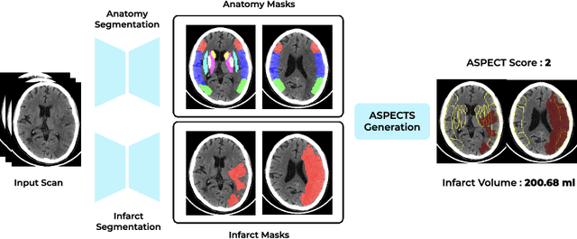

Abstract:A stroke occurs when an artery in the brain ruptures and bleeds or when the blood supply to the brain is cut off. Blood and oxygen cannot reach the brain's tissues due to the rupture or obstruction resulting in tissue death. The Middle cerebral artery (MCA) is the largest cerebral artery and the most commonly damaged vessel in stroke. The quick onset of a focused neurological deficit caused by interruption of blood flow in the territory supplied by the MCA is known as an MCA stroke. Alberta stroke programme early CT score (ASPECTS) is used to estimate the extent of early ischemic changes in patients with MCA stroke. This study proposes a deep learning-based method to score the CT scan for ASPECTS. Our work has three highlights. First, we propose a novel method for medical image segmentation for stroke detection. Second, we show the effectiveness of AI solution for fully-automated ASPECT scoring with reduced diagnosis time for a given non-contrast CT (NCCT) Scan. Our algorithms show a dice similarity coefficient of 0.64 for the MCA anatomy segmentation and 0.72 for the infarcts segmentation. Lastly, we show that our model's performance is inline with inter-reader variability between radiologists.
Development and Validation of Deep Learning Algorithms for Detection of Critical Findings in Head CT Scans
Apr 12, 2018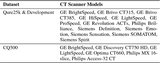
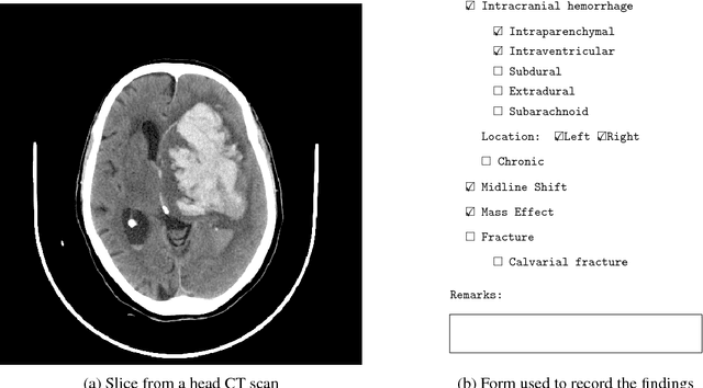
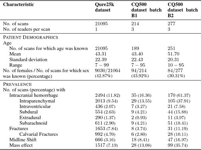
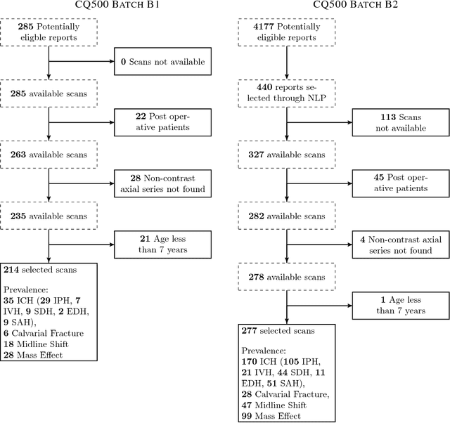
Abstract:Importance: Non-contrast head CT scan is the current standard for initial imaging of patients with head trauma or stroke symptoms. Objective: To develop and validate a set of deep learning algorithms for automated detection of following key findings from non-contrast head CT scans: intracranial hemorrhage (ICH) and its types, intraparenchymal (IPH), intraventricular (IVH), subdural (SDH), extradural (EDH) and subarachnoid (SAH) hemorrhages, calvarial fractures, midline shift and mass effect. Design and Settings: We retrospectively collected a dataset containing 313,318 head CT scans along with their clinical reports from various centers. A part of this dataset (Qure25k dataset) was used to validate and the rest to develop algorithms. Additionally, a dataset (CQ500 dataset) was collected from different centers in two batches B1 & B2 to clinically validate the algorithms. Main Outcomes and Measures: Original clinical radiology report and consensus of three independent radiologists were considered as gold standard for Qure25k and CQ500 datasets respectively. Area under receiver operating characteristics curve (AUC) for each finding was primarily used to evaluate the algorithms. Results: Qure25k dataset contained 21,095 scans (mean age 43.31; 42.87% female) while batches B1 and B2 of CQ500 dataset consisted of 214 (mean age 43.40; 43.92% female) and 277 (mean age 51.70; 30.31% female) scans respectively. On Qure25k dataset, the algorithms achieved AUCs of 0.9194, 0.8977, 0.9559, 0.9161, 0.9288 and 0.9044 for detecting ICH, IPH, IVH, SDH, EDH and SAH respectively. AUCs for the same on CQ500 dataset were 0.9419, 0.9544, 0.9310, 0.9521, 0.9731 and 0.9574 respectively. For detecting calvarial fractures, midline shift and mass effect, AUCs on Qure25k dataset were 0.9244, 0.9276 and 0.8583 respectively, while AUCs on CQ500 dataset were 0.9624, 0.9697 and 0.9216 respectively.
 Add to Chrome
 Add to Firefox
 Add to Edge