 Add to Chrome
 Add to Firefox
 Add to Edge
Harmonizing MR Images Across 100+ Scanners: Multi-site Validation with Traveling Subjects and Real-world Protocols
Apr 21, 2026Reliable harmonization of heterogeneous magnetic resonance~(MR) image datasets, especially those acquired in pragmatic clinical trials, is critical to advance multi-center neuroimaging studies and translational machine learning in healthcare. We present an enhanced and rigorously validated version of the HACA3 harmonization algorithm, which we refer to as HACA3$^+$, incorporating key methodological enhancements: (1)~an improved artifact encoder to better isolate and mitigate image artifacts, (2)~background and foreground-sensitive attention mechanisms to increase harmonization specificity, and (3)~extensive training using data spanning 100+ scanners from 64 independent sites, providing a broader diversity of scanners than other harmonization methods. Our study focuses on four commonly acquired MR image contrasts (T1-weighted, T2-weighted, proton density, \& fluid-attenuated inversion recovery), reflecting realistic clinical protocols. We perform inter-site harmonization experiments using traveling subjects to assess the generalization and robustness of the harmonization model. We compare the results of the publicly available version of HACA3 and our implementation, HACA3$^+$. Downstream relevance is further established through whole brain segmentation and image imputation. Finally, we justify each enhancement through an ablation experiment. Pre-trained weights and code for HACA3$^+$ are made publicly available at https://github.com/shays15/haca3-plus.
MSRepaint: Multiple Sclerosis Repaint with Conditional Denoising Diffusion Implicit Model for Bidirectional Lesion Filling and Synthesis
Oct 02, 2025In multiple sclerosis, lesions interfere with automated magnetic resonance imaging analyses such as brain parcellation and deformable registration, while lesion segmentation models are hindered by the limited availability of annotated training data. To address both issues, we propose MSRepaint, a unified diffusion-based generative model for bidirectional lesion filling and synthesis that restores anatomical continuity for downstream analyses and augments segmentation through realistic data generation. MSRepaint conditions on spatial lesion masks for voxel-level control, incorporates contrast dropout to handle missing inputs, integrates a repainting mechanism to preserve surrounding anatomy during lesion filling and synthesis, and employs a multi-view DDIM inversion and fusion pipeline for 3D consistency with fast inference. Extensive evaluations demonstrate the effectiveness of MSRepaint across multiple tasks. For lesion filling, we evaluate both the accuracy within the filled regions and the impact on downstream tasks including brain parcellation and deformable registration. MSRepaint outperforms the traditional lesion filling methods FSL and NiftySeg, and achieves accuracy on par with FastSurfer-LIT, a recent diffusion model-based inpainting method, while offering over 20 times faster inference. For lesion synthesis, state-of-the-art MS lesion segmentation models trained on MSRepaint-synthesized data outperform those trained on CarveMix-synthesized data or real ISBI challenge training data across multiple benchmarks, including the MICCAI 2016 and UMCL datasets. Additionally, we demonstrate that MSRepaint's unified bidirectional filling and synthesis capability, with full spatial control over lesion appearance, enables high-fidelity simulation of lesion evolution in longitudinal MS progression.
Topology guaranteed segmentation of the human retina from OCT using convolutional neural networks
Mar 14, 2018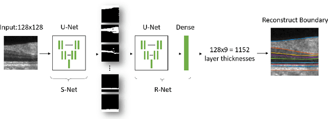
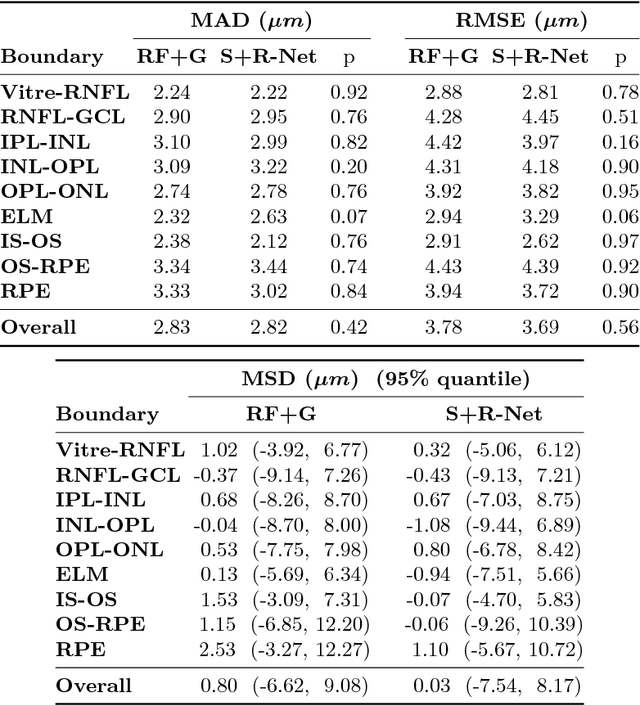
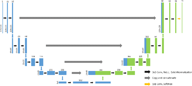
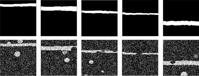
Optical coherence tomography (OCT) is a noninvasive imaging modality which can be used to obtain depth images of the retina. The changing layer thicknesses can thus be quantified by analyzing these OCT images, moreover these changes have been shown to correlate with disease progression in multiple sclerosis. Recent automated retinal layer segmentation tools use machine learning methods to perform pixel-wise labeling and graph methods to guarantee the layer hierarchy or topology. However, graph parameters like distance and smoothness constraints must be experimentally assigned by retinal region and pathology, thus degrading the flexibility and time efficiency of the whole framework. In this paper, we develop cascaded deep networks to provide a topologically correct segmentation of the retinal layers in a single feed forward propagation. The first network (S-Net) performs pixel-wise labeling and the second regression network (R-Net) takes the topologically unconstrained S-Net results and outputs layer thicknesses for each layer and each position. Relu activation is used as the final operation of the R-Net which guarantees non-negativity of the output layer thickness. Since the segmentation boundary position is acquired by summing up the corresponding non-negative layer thicknesses, the layer ordering (i.e., topology) of the reconstructed boundaries is guaranteed even at the fovea where the distances between boundaries can be zero. The R-Net is trained using simulated masks and thus can be generalized to provide topology guaranteed segmentation for other layered structures. This deep network has achieved comparable mean absolute boundary error (2.82 {\mu}m) to state-of-the-art graph methods (2.83 {\mu}m).