Se-Hong Oh
So You Want to Image Myelin Using MRI: Advanced Magnetic Susceptibility Imaging for Myelin
Jan 08, 2024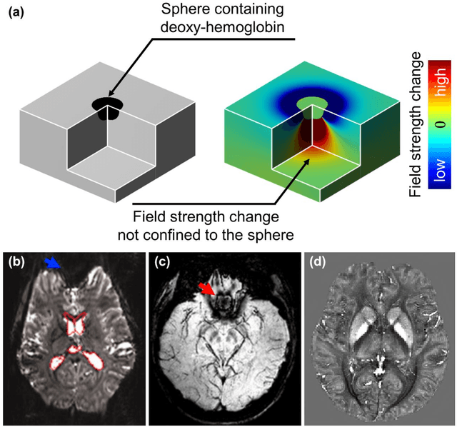

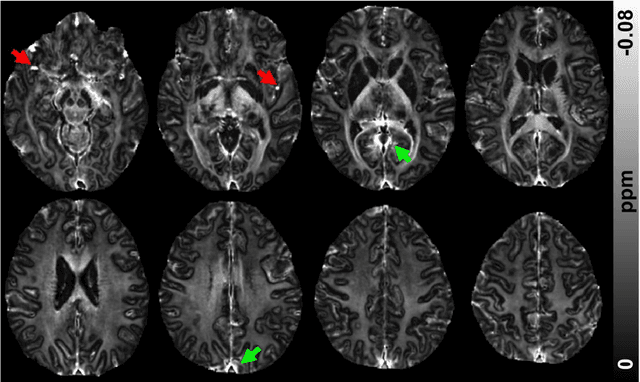
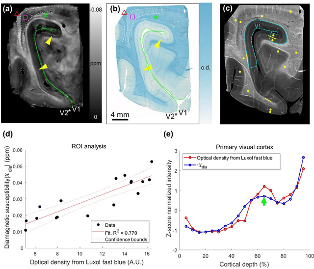
Abstract:In MRI, researchers have long endeavored to effectively visualize myelin distribution in the brain, a pursuit with significant implications for both scientific research and clinical applications. Over time, various methods such as myelin water imaging, magnetization transfer imaging, and relaxometric imaging have been developed, each carrying distinct advantages and limitations. Recently, an innovative technique named as magnetic susceptibility source separation has emerged, introducing a novel surrogate biomarker for myelin in the form of a diamagnetic susceptibility map. This paper comprehensively reviews this cutting-edge method, providing the fundamental concepts of magnetic susceptibility, susceptibility imaging, and the validation of the diamagnetic susceptibility map as a myelin biomarker. Additionally, the paper explores essential aspects of data acquisition and processing, offering practical insights for readers. A comparison with established myelin imaging methods is also presented, and both current and prospective clinical and scientific applications are discussed to provide a holistic understanding of the technique. This work aims to serve as a foundational resource for newcomers entering this dynamic and rapidly expanding field.
DIFFnet: Diffusion parameter mapping network generalized for input diffusion gradient schemes and bvalues
Feb 04, 2021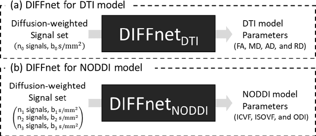
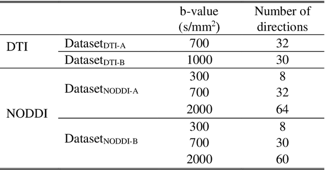
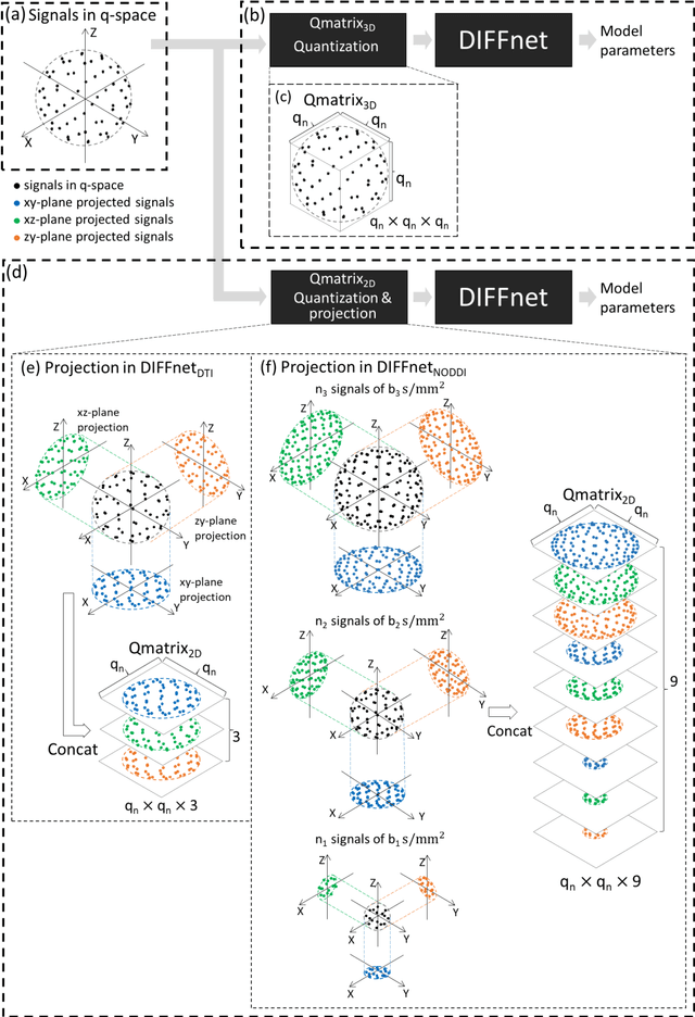
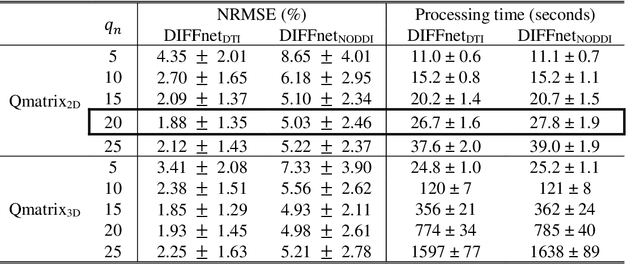
Abstract:In MRI, deep neural networks have been proposed to reconstruct diffusion model parameters. However, the inputs of the networks were designed for a specific diffusion gradient scheme (i.e., diffusion gradient directions and numbers) and a specific b-value that are the same as the training data. In this study, a new deep neural network, referred to as DIFFnet, is developed to function as a generalized reconstruction tool of the diffusion-weighted signals for various gradient schemes and b-values. For generalization, diffusion signals are normalized in a q-space and then projected and quantized, producing a matrix (Qmatrix) as an input for the network. To demonstrate the validity of this approach, DIFFnet is evaluated for diffusion tensor imaging (DIFFnetDTI) and for neurite orientation dispersion and density imaging (DIFFnetNODDI). In each model, two datasets with different gradient schemes and b-values are tested. The results demonstrate accurate reconstruction of the diffusion parameters at substantially reduced processing time (approximately 8.7 times and 2240 times faster processing time than conventional methods in DTI and NODDI, respectively; less than 4% mean normalized root-mean-square errors (NRMSE) in DTI and less than 8% in NODDI). The generalization capability of the networks was further validated using reduced numbers of diffusion signals from the datasets. Different from previously proposed deep neural networks, DIFFnet does not require any specific gradient scheme and b-value for its input. As a result, it can be adopted as an online reconstruction tool for various complex diffusion imaging.
Deep Reinforcement Learning Designed RF Pulse: $DeepRF_{SLR}$
Dec 19, 2019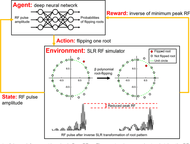
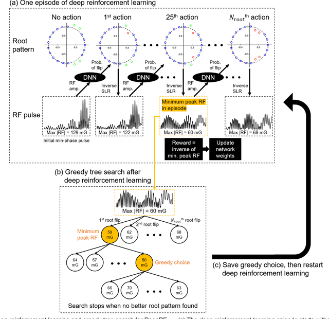
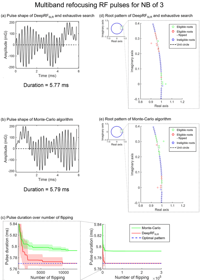
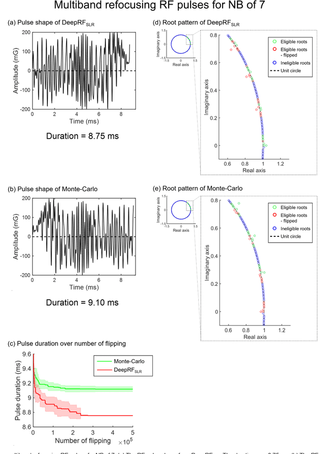
Abstract:A novel approach of applying deep reinforcement learning to an RF pulse design is introduced. This method, which is referred to as $DeepRF_{SLR}$, is designed to minimize the peak amplitude or, equivalently, minimize the pulse duration of a multiband refocusing pulse generated by the Shinar Le-Roux (SLR) algorithm. In the method, the root pattern of SLR polynomial, which determines the RF pulse shape, is optimized by iterative applications of deep reinforcement learning and greedy tree search. When tested for the designs of the multiband factors of three and seven RFs, $DeepRF_{SLR}$ demonstrated improved performance compared to conventional methods, generating shorter duration RF pulses in shorter computational time. In the experiments, the RF pulse from $DeepRF_{SLR}$ produced a slice profile similar to the minimum-phase SLR RF pulse and the profiles matched to that of the computer simulation. Our approach suggests a new way of designing an RF by applying a machine learning algorithm, demonstrating a machine-designed MRI sequence.
 Add to Chrome
 Add to Firefox
 Add to Edge