Saroj Jayasinghe
LiverUSRecon: Automatic 3D Reconstruction and Volumetry of the Liver with a Few Partial Ultrasound Scans
Jun 27, 2024Abstract:3D reconstruction of the liver for volumetry is important for qualitative analysis and disease diagnosis. Liver volumetry using ultrasound (US) scans, although advantageous due to less acquisition time and safety, is challenging due to the inherent noisiness in US scans, blurry boundaries, and partial liver visibility. We address these challenges by using the segmentation masks of a few incomplete sagittal-plane US scans of the liver in conjunction with a statistical shape model (SSM) built using a set of CT scans of the liver. We compute the shape parameters needed to warp this canonical SSM to fit the US scans through a parametric regression network. The resulting 3D liver reconstruction is accurate and leads to automatic liver volume calculation. We evaluate the accuracy of the estimated liver volumes with respect to CT segmentation volumes using RMSE. Our volume computation is statistically much closer to the volume estimated using CT scans than the volume computed using Childs' method by radiologists: p-value of 0.094 (>0.05) says that there is no significant difference between CT segmentation volumes and ours in contrast to Childs' method. We validate our method using investigations (ablation studies) on the US image resolution, the number of CT scans used for SSM, the number of principal components, and the number of input US scans. To the best of our knowledge, this is the first automatic liver volumetry system using a few incomplete US scans given a set of CT scans of livers for SSM.
A Thickness Sensitive Vessel Extraction Framework for Retinal and Conjunctival Vascular Tortuosity Analysis
Jan 02, 2021


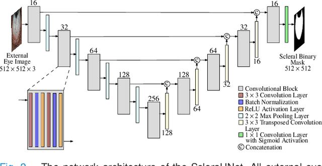
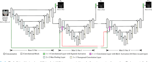
Abstract:Systemic diseases such as diabetes, hypertension, atherosclerosis are among the leading causes of annual human mortality rate. It is suggested that retinal and conjunctival vascular tortuosity is a potential biomarker for such systemic diseases. Most importantly, it is observed that the tortuosity depends on the thickness of these vessels. Therefore, selective calculation of tortuosity within specific vessel thicknesses is required depending on the disease being analysed. In this paper, we propose a thickness sensitive vessel extraction framework that is primarily applicable for studies related to retinal and conjunctival vascular tortuosity. The framework uses a Convolutional Neural Network based on the IterNet architecture to obtain probability maps of the entire vasculature. They are then processed by a multi-scale vessel enhancement technique that exploits both fine and coarse structural vascular details of these probability maps in order to extract vessels of specified thicknesses. We evaluated the proposed framework on four datasets including DRIVE and SBVPI, and obtained Matthew's Correlation Coefficient values greater than 0.71 for all the datasets. In addition, the proposed framework was utilized to determine the association of diabetes with retinal and conjunctival vascular tortuosity. We observed that retinal vascular tortuosity (Eccentricity based Tortuosity Index) of the diabetic group was significantly higher (p < .05) than that of the non-diabetic group and that conjunctival vascular tortuosity (Total Curvature normalized by Arc Length) of diabetic group was significantly lower (p < .05) than that of the non-diabetic group. These observations were in agreement with the literature, strengthening the suitability of the proposed framework.
 Add to Chrome
 Add to Firefox
 Add to Edge