Sang Jun Park
Generation of Structurally Realistic Retinal Fundus Images with Diffusion Models
May 11, 2023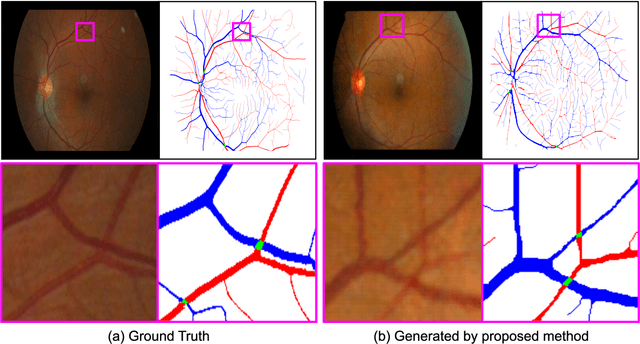
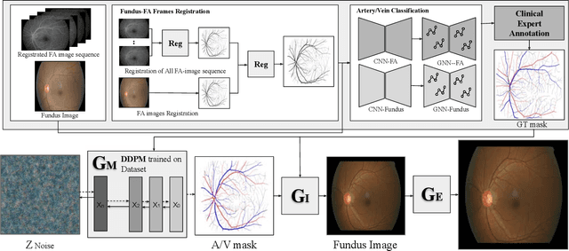
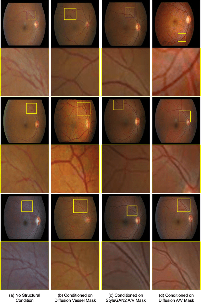
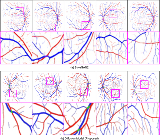
Abstract:We introduce a new technique for generating retinal fundus images that have anatomically accurate vascular structures, using diffusion models. We generate artery/vein masks to create the vascular structure, which we then condition to produce retinal fundus images. The proposed method can generate high-quality images with more realistic vascular structures and can create a diverse range of images based on the strengths of the diffusion model. We present quantitative evaluations that demonstrate the performance improvement using our method for data augmentation on vessel segmentation and artery/vein classification. We also present Turing test results by clinical experts, showing that our generated images are difficult to distinguish with real images. We believe that our method can be applied to construct stand-alone datasets that are irrelevant of patient privacy.
Classification of Findings with Localized Lesions in Fundoscopic Images using a Regionally Guided CNN
Nov 02, 2018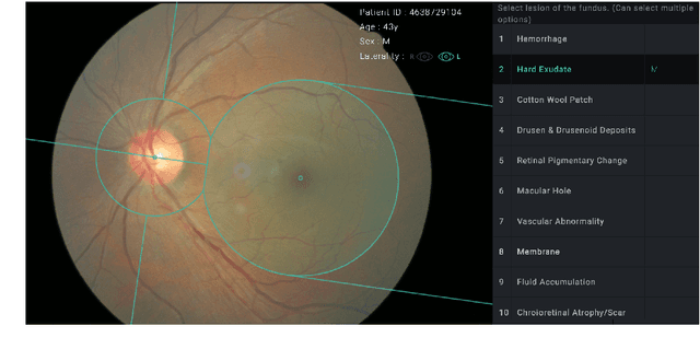

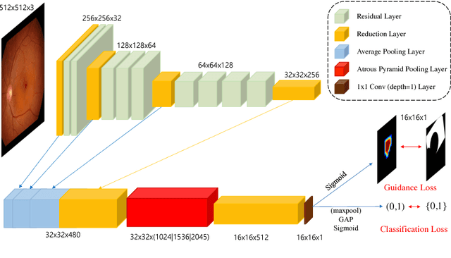
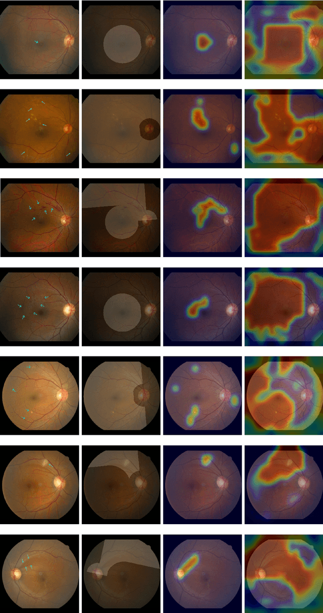
Abstract:Fundoscopic images are often investigated by ophthalmologists to spot abnormal lesions to make diagnoses. Recent successes of convolutional neural networks are confined to diagnoses of few diseases without proper localization of lesion. In this paper, we propose an efficient annotation method for localizing lesions and a CNN architecture that can classify an individual finding and localize the lesions at the same time. Also, we introduce a new loss function to guide the network to learn meaningful patterns with the guidance of the regional annotations. In experiments, we demonstrate that our network performed better than the widely used network and the guidance loss helps achieve higher AUROC up to 4.1% and superior localization capability.
Scale Space Approximation in Convolutional Neural Networks for Retinal Vessel Segmentation
Oct 18, 2018
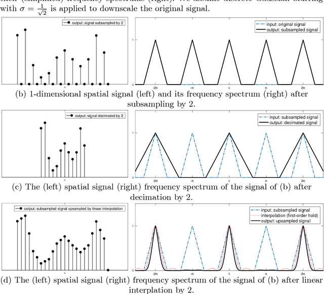
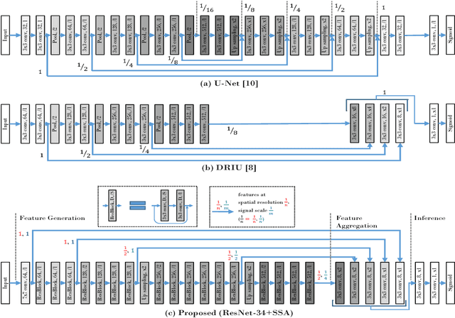
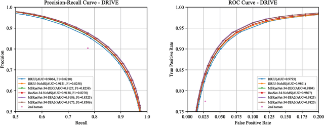
Abstract:Retinal images have the highest resolution and clarity among medical images. Thus, vessel analysis in retinal images may facilitate early diagnosis and treatment of many chronic diseases. In this paper, we propose a novel multi-scale residual convolutional neural network structure based on a \emph{scale-space approximation (SSA)} block of layers, comprising subsampling and subsequent upsampling, for multi-scale representation. Through analysis in the frequency domain, we show that this block structure is a close approximation of Gaussian filtering, the operation to achieve scale variations in scale-space theory. Experimental evaluations demonstrate that the proposed network outperforms current state-of-the-art methods. Ablative analysis shows that the SSA is indeed an important factor in performance improvement.
Retinal Vessel Segmentation in Fundoscopic Images with Generative Adversarial Networks
Jun 28, 2017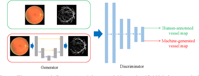

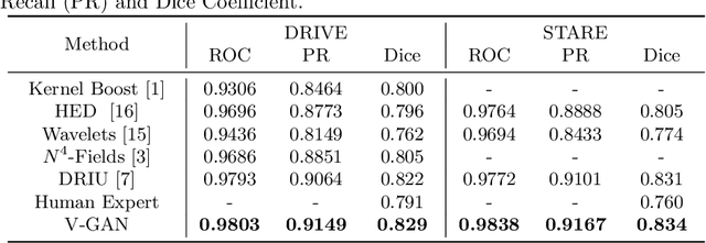
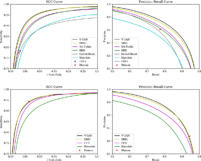
Abstract:Retinal vessel segmentation is an indispensable step for automatic detection of retinal diseases with fundoscopic images. Though many approaches have been proposed, existing methods tend to miss fine vessels or allow false positives at terminal branches. Let alone under-segmentation, over-segmentation is also problematic when quantitative studies need to measure the precise width of vessels. In this paper, we present a method that generates the precise map of retinal vessels using generative adversarial training. Our methods achieve dice coefficient of 0.829 on DRIVE dataset and 0.834 on STARE dataset which is the state-of-the-art performance on both datasets.
 Add to Chrome
 Add to Firefox
 Add to Edge