Sahar Shariflou
RVD: A Handheld Device-Based Fundus Video Dataset for Retinal Vessel Segmentation
Jul 13, 2023Abstract:Retinal vessel segmentation is generally grounded in image-based datasets collected with bench-top devices. The static images naturally lose the dynamic characteristics of retina fluctuation, resulting in diminished dataset richness, and the usage of bench-top devices further restricts dataset scalability due to its limited accessibility. Considering these limitations, we introduce the first video-based retinal dataset by employing handheld devices for data acquisition. The dataset comprises 635 smartphone-based fundus videos collected from four different clinics, involving 415 patients from 50 to 75 years old. It delivers comprehensive and precise annotations of retinal structures in both spatial and temporal dimensions, aiming to advance the landscape of vasculature segmentation. Specifically, the dataset provides three levels of spatial annotations: binary vessel masks for overall retinal structure delineation, general vein-artery masks for distinguishing the vein and artery, and fine-grained vein-artery masks for further characterizing the granularities of each artery and vein. In addition, the dataset offers temporal annotations that capture the vessel pulsation characteristics, assisting in detecting ocular diseases that require fine-grained recognition of hemodynamic fluctuation. In application, our dataset exhibits a significant domain shift with respect to data captured by bench-top devices, thus posing great challenges to existing methods. In the experiments, we provide evaluation metrics and benchmark results on our dataset, reflecting both the potential and challenges it offers for vessel segmentation tasks. We hope this challenging dataset would significantly contribute to the development of eye disease diagnosis and early prevention.
Autonomous Stabilization of Retinal Videos for Streamlining Assessment of Spontaneous Venous Pulsations
May 10, 2023


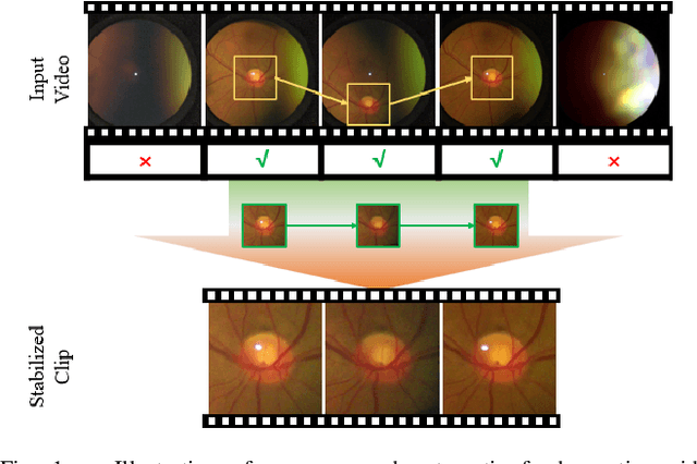
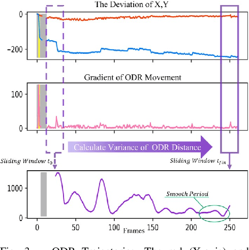
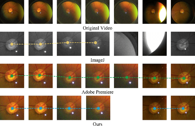
Abstract:Spontaneous retinal Venous Pulsations (SVP) are rhythmic changes in the caliber of the central retinal vein and are observed in the optic disc region (ODR) of the retina. Its absence is a critical indicator of various ocular or neurological abnormalities. Recent advances in imaging technology have enabled the development of portable smartphone-based devices for observing the retina and assessment of SVPs. However, the quality of smartphone-based retinal videos is often poor due to noise and image jitting, which in return, can severely obstruct the observation of SVPs. In this work, we developed a fully automated retinal video stabilization method that enables the examination of SVPs captured by various mobile devices. Specifically, we first propose an ODR Spatio-Temporal Localization (ODR-STL) module to localize visible ODR and remove noisy and jittering frames. Then, we introduce a Noise-Aware Template Matching (NATM) module to stabilize high-quality video segments at a fixed position in the field of view. After the processing, the SVPs can be easily observed in the stabilized videos, significantly facilitating user observations. Furthermore, our method is cost-effective and has been tested in both subjective and objective evaluations. Both of the evaluations support its effectiveness in facilitating the observation of SVPs. This can improve the timely diagnosis and treatment of associated diseases, making it a valuable tool for eye health professionals.
 Add to Chrome
 Add to Firefox
 Add to Edge