Piyamate Wisanuvej
SurReal: enhancing Surgical simulation Realism using style transfer
Nov 07, 2018


Abstract:Surgical simulation is an increasingly important element of surgical education. Using simulation can be a means to address some of the significant challenges in developing surgical skills with limited time and resources. The photo-realistic fidelity of simulations is a key feature that can improve the experience and transfer ratio of trainees. In this paper, we demonstrate how we can enhance the visual fidelity of existing surgical simulation by performing style transfer of multi-class labels from real surgical video onto synthetic content. We demonstrate our approach on simulations of cataract surgery using real data labels from an existing public dataset. Our results highlight the feasibility of the approach and also the powerful possibility to extend this technique to incorporate additional temporal constraints and to different applications.
Intraoperative robotic-assisted large-area high-speed microscopic imaging and intervention
Aug 13, 2018


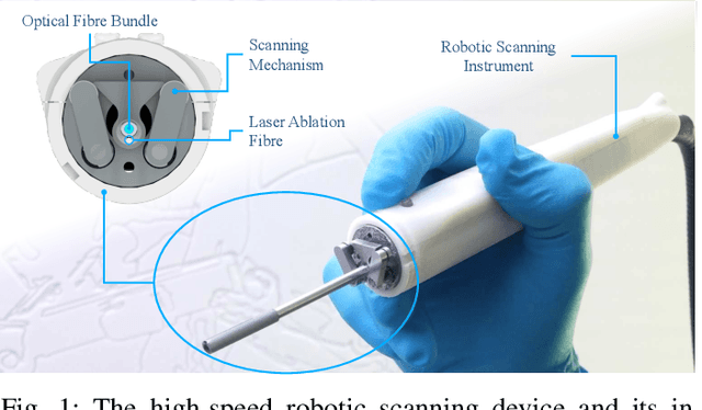
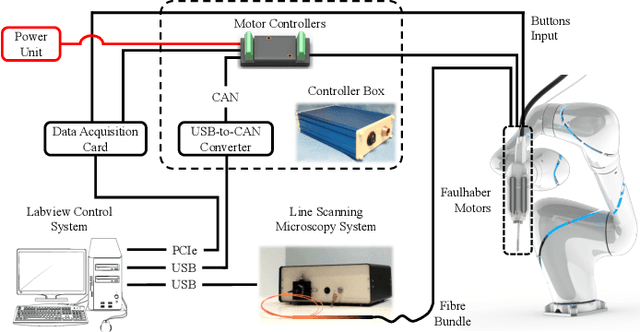
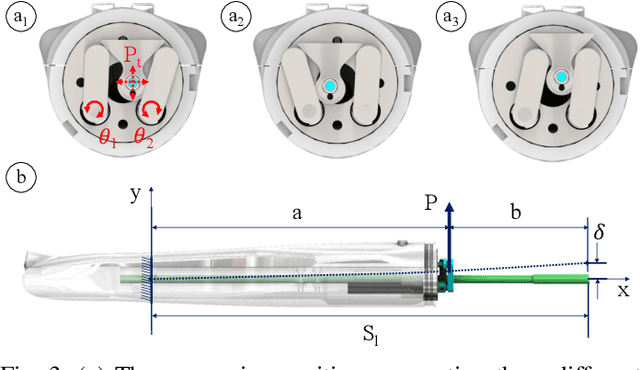
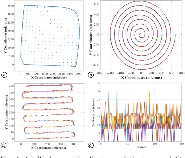
Abstract:Objective: Probe-based confocal endomicroscopy is an emerging high-magnification optical imaging technique that provides in vivo and in situ cellular-level imaging for real-time assessment of tissue pathology. Endomicroscopy could potentially be used for intraoperative surgical guidance, but it is challenging to assess a surgical site using individual microscopic images due to the limited field-of-view and difficulties associated with manually manipulating the probe. Methods: In this paper, a novel robotic device for large-area endomicroscopy imaging is proposed, demonstrating a rapid, but highly accurate, scanning mechanism with image-based motion control which is able to generate histology-like endomicroscopy mosaics. The device also includes, for the first time in robotic-assisted endomicroscopy, the capability to ablate tissue without the need for an additional tool. Results: The device achieves pre-programmed trajectories with positioning accuracy of less than 30 um, while the image-based approach demonstrated that it can suppress random motion disturbances up to 1.25 mm/s. Mosaics are presented from a range of ex vivo human and animal tissues, over areas of more than 3 mm^2, scanned in approximate 10 seconds. Conclusion: This work demonstrates the potential of the proposed instrument to generate large-area, high-resolution microscopic images for intraoperative tissue identification and margin assessment. Significance: This approach presents an important alternative to current histology techniques, significantly reducing the tissue assessment time, while simultaneously providing the capability to mark and ablate suspicious areas intraoperatively.
Affordable Mobile-based Simulator for Robotic Surgery
Jul 20, 2018


Abstract:Robotic surgery and novel surgical instrumentation present great potentials towards safer, more accurate and consistent minimally invasive surgery. However, their adoption is dependent to the access to training facilities and extensive surgical training. Robotic instruments require different dexterity skills compared to open or laparoscopic. Surgeons, therefore, are required to invest significant time by attending extensive training programs. Contrary, hands on experiences represent an additional operational cost for hospitals as the availability of robotic systems for training purposes is limited. All these technological and financial barriers for surgeons and hospitals hinder the adoption of robotic surgery. In this paper, we present a mobile dexterity training kit to develop basic surgical techniques within an affordable setting. The system could be used to train basic surgical gestures and to develop the motor skills needed for manoeuvring robotic instruments. Our work presents the architecture and components needed to create a simulated environment for training sub-tasks as well as a design for portable mobile manipulators that can be used as master controllers of different instruments. A preliminary study results demonstrate usability and skills development with this system.
 Add to Chrome
 Add to Firefox
 Add to Edge