Peter Gatehouse
HDL: Hybrid Deep Learning for the Synthesis of Myocardial Velocity Maps in Digital Twins for Cardiac Analysis
Mar 09, 2022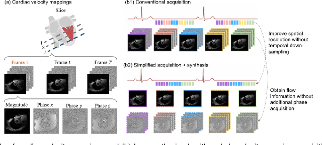
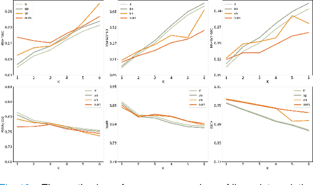
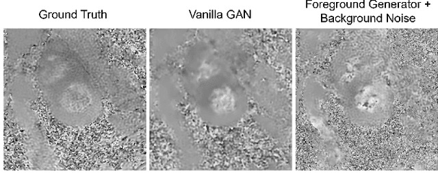
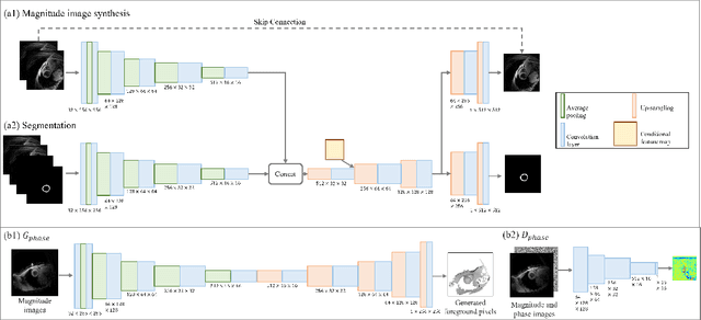
Abstract:Synthetic digital twins based on medical data accelerate the acquisition, labelling and decision making procedure in digital healthcare. A core part of digital healthcare twins is model-based data synthesis, which permits the generation of realistic medical signals without requiring to cope with the modelling complexity of anatomical and biochemical phenomena producing them in reality. Unfortunately, algorithms for cardiac data synthesis have been so far scarcely studied in the literature. An important imaging modality in the cardiac examination is three-directional CINE multi-slice myocardial velocity mapping (3Dir MVM), which provides a quantitative assessment of cardiac motion in three orthogonal directions of the left ventricle. The long acquisition time and complex acquisition produce make it more urgent to produce synthetic digital twins of this imaging modality. In this study, we propose a hybrid deep learning (HDL) network, especially for synthetic 3Dir MVM data. Our algorithm is featured by a hybrid UNet and a Generative Adversarial Network with a foreground-background generation scheme. The experimental results show that from temporally down-sampled magnitude CINE images (six times), our proposed algorithm can still successfully synthesise high temporal resolution 3Dir MVM CMR data (PSNR=42.32) with precise left ventricle segmentation (DICE=0.92). These performance scores indicate that our proposed HDL algorithm can be implemented in real-world digital twins for myocardial velocity mapping data simulation. To the best of our knowledge, this work is the first one in the literature investigating digital twins of the 3Dir MVM CMR, which has shown great potential for improving the efficiency of clinical studies via synthesised cardiac data.
Synthetic Velocity Mapping Cardiac MRI Coupled with Automated Left Ventricle Segmentation
Oct 04, 2021

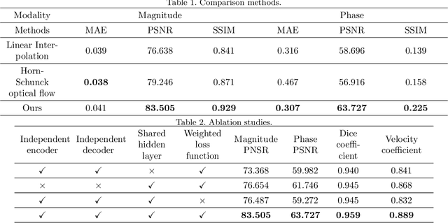

Abstract:Temporal patterns of cardiac motion provide important information for cardiac disease diagnosis. This pattern could be obtained by three-directional CINE multi-slice left ventricular myocardial velocity mapping (3Dir MVM), which is a cardiac MR technique providing magnitude and phase information of the myocardial motion simultaneously. However, long acquisition time limits the usage of this technique by causing breathing artifacts, while shortening the time causes low temporal resolution and may provide an inaccurate assessment of cardiac motion. In this study, we proposed a frame synthesis algorithm to increase the temporal resolution of 3Dir MVM data. Our algorithm is featured by 1) three attention-based encoders which accept magnitude images, phase images, and myocardium segmentation masks respectively as inputs; 2) three decoders that output the interpolated frames and corresponding myocardium segmentation results; and 3) loss functions highlighting myocardium pixels. Our algorithm can not only increase the temporal resolution 3Dir MVMs, but can also generates the myocardium segmentation results at the same time.
Three-Dimensional Embedded Attentive RNN (3D-EAR) Segmentor for Left Ventricle Delineation from Myocardial Velocity Mapping
Apr 26, 2021

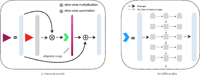

Abstract:Myocardial Velocity Mapping Cardiac MR (MVM-CMR) can be used to measure global and regional myocardial velocities with proved reproducibility. Accurate left ventricle delineation is a prerequisite for robust and reproducible myocardial velocity estimation. Conventional manual segmentation on this dataset can be time-consuming and subjective, and an effective fully automated delineation method is highly in demand. By leveraging recently proposed deep learning-based semantic segmentation approaches, in this study, we propose a novel fully automated framework incorporating a 3D-UNet backbone architecture with Embedded multichannel Attention mechanism and LSTM based Recurrent neural networks (RNN) for the MVM-CMR datasets (dubbed 3D-EAR segmentor). The proposed method also utilises the amalgamation of magnitude and phase images as input to realise an information fusion of this multichannel dataset and exploring the correlations of temporal frames via the embedded RNN. By comparing the baseline model of 3D-UNet and ablation studies with and without embedded attentive LSTM modules and various loss functions, we can demonstrate that the proposed model has outperformed the state-of-the-art baseline models with significant improvement.
Automated Multi-Channel Segmentation for the 4D Myocardial Velocity Mapping Cardiac MR
Dec 16, 2020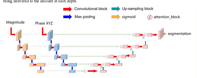


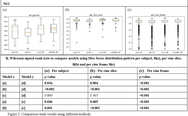
Abstract:Four-dimensional (4D) left ventricular myocardial velocity mapping (MVM) is a cardiac magnetic resonance (CMR) technique that allows assessment of cardiac motion in three orthogonal directions. Accurate and reproducible delineation of the myocardium is crucial for accurate analysis of peak systolic and diastolic myocardial velocities. In addition to the conventionally available magnitude CMR data, 4D MVM also acquires three velocity-encoded phase datasets which are used to generate velocity maps. These can be used to facilitate and improve myocardial delineation. Based on the success of deep learning in medical image processing, we propose a novel automated framework that improves the standard U-Net based methods on these CMR multi-channel data (magnitude and phase) by cross-channel fusion with attention module and shape information based post-processing to achieve accurate delineation of both epicardium and endocardium contours. To evaluate the results, we employ the widely used Dice scores and the quantification of myocardial longitudinal peak velocities. Our proposed network trained with multi-channel data shows enhanced performance compared to standard U-Net based networks trained with single-channel data. Based on the results, our method provides compelling evidence for the design and application for the multi-channel image analysis of the 4D MVM CMR data.
 Add to Chrome
 Add to Firefox
 Add to Edge