Olivier Rukundo
Beyond Nearest Neighbor Interpolation in Data Augmentation
Apr 02, 2025Abstract:Avoiding the risk of undefined categorical labels using nearest neighbor interpolation overlooks the risk of exacerbating pixel level annotation errors in data augmentation. To simultaneously avoid these risks, the author modified convolutional neural networks data transformation functions by incorporating a modified geometric transformation function to improve the quality of augmented data by removing the reliance on nearest neighbor interpolation and integrating a mean based class filtering mechanism to handle undefined categorical labels with alternative interpolation algorithms. Experiments on semantic segmentation tasks using three medical image datasets demonstrated both qualitative and quantitative improvements with alternative interpolation algorithms.
Convolutional Neural Networks for Automatic Detection of Intact Adenovirus from TEM Imaging with Debris, Broken and Artefacts Particles
Nov 06, 2023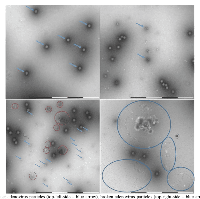
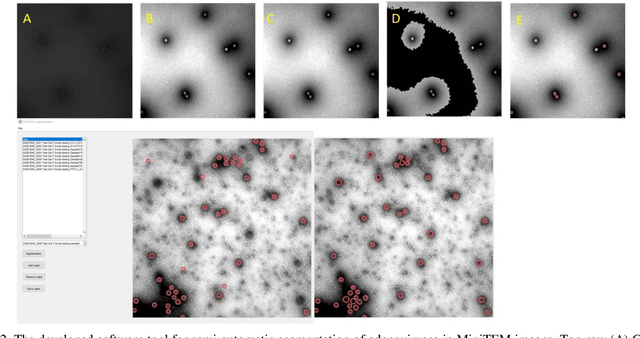
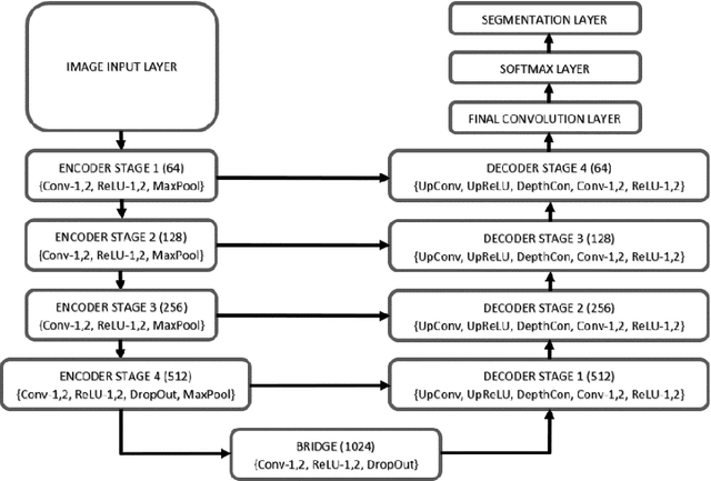
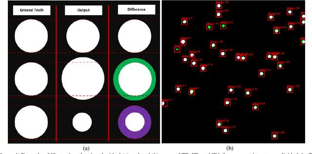
Abstract:Regular monitoring of the primary particles and purity profiles of a drug product during development and manufacturing processes is essential for manufacturers to avoid product variability and contamination. Transmission electron microscopy (TEM) imaging helps manufacturers predict how changes affect particle characteristics and purity for virus-based gene therapy vector products and intermediates. Since intact particles can characterize efficacious products, it is beneficial to automate the detection of intact adenovirus against a non-intact-viral background mixed with debris, broken, and artefact particles. In the presence of such particles, detecting intact adenoviruses becomes more challenging. To overcome the challenge, due to such a presence, we developed a software tool for semi-automatic annotation and segmentation of adenoviruses and a software tool for automatic segmentation and detection of intact adenoviruses in TEM imaging systems. The developed semi-automatic tool exploited conventional image analysis techniques while the automatic tool was built based on convolutional neural networks and image analysis techniques. Our quantitative and qualitative evaluations showed outstanding true positive detection rates compared to false positive and negative rates where adenoviruses were nicely detected without mistaking them for real debris, broken adenoviruses, and/or staining artefacts.
Evaluation of Extra Pixel Interpolation with Mask Processing for Medical Image Segmentation with Deep Learning
Feb 22, 2023Abstract:In this study, the author evaluated the use of an extra pixel interpolation algorithm with mask processing versus non-extra pixel interpolation algorithm when interpolating training dataset images and masks for medical image segmentation with deep learning. The author also examined scenarios of interpolating dataset images and masks using different algorithms: extra pixel for interpolating dataset images and non-extra pixel for interpolating dataset masks. The evaluation outcomes revealed that training on datasets consisting of images and masks both interpolated using the extra pixel bicubic interpolation (BIC) resulted in better segmentation accuracy compared to using either the non-extra pixel nearest neighbor interpolation (NN) or BIC for dataset images and NN for dataset masks. Specifically, the evaluation revealed that the BIC-BIC network was a 8.9578 % (with image size 256 x 256) and a 1.0496 % (with image size 384 x 384) increase of NN-NN network compared to the NN-BIC network which was a 8.3127 % (with image size 256 x 256) and a 0.2887 % (with image size 384 x 384) increase of NN-NN network.
Stochastic Rounding for Image Interpolation and Scan Conversion
Oct 25, 2021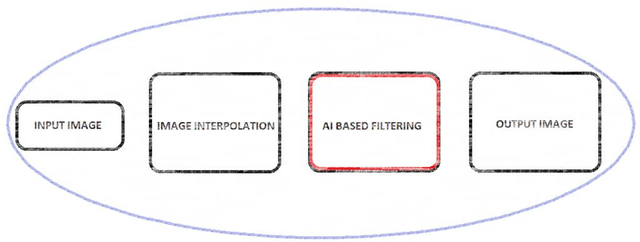
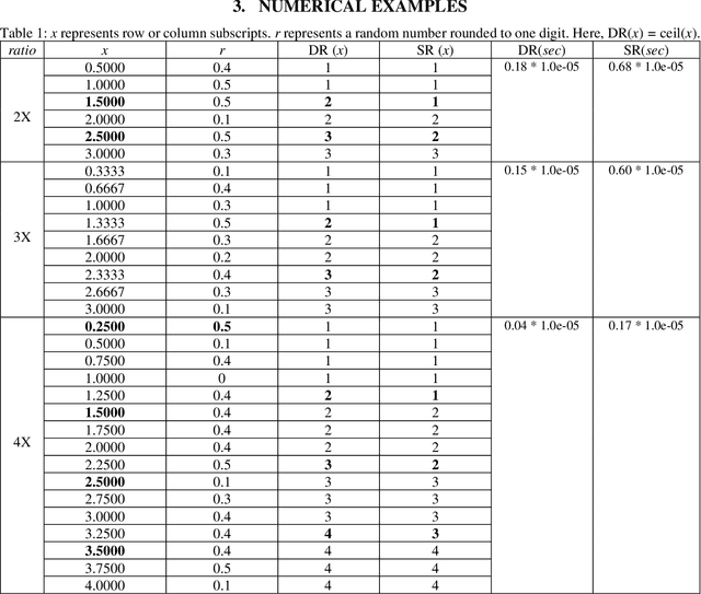
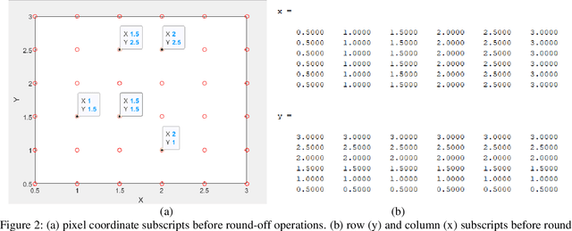

Abstract:The stochastic rounding (SR) function is introduced to demonstrate the effects of stochastically rounding row and column subscripts on image interpolation quality in nearest neighbor interpolation (NNI). The introduced SR function is based on a pseudorandom number that enables the pseudorandom rounding up or down of any non-integer row and column subscripts. Also, the SR function exceptionally enables rounding up of any possible cases of subscript inputs that are inferior to a pseudorandom number - especially at a high interpolation scaling ratio. The quality of NNI-SR interpolated images is evaluated against the quality of reference images - before and after applying smoothing and sharpening filters, mentioned. The quality of NNI-SR interpolated scan conversion video frames is evaluated without using any references - focusing on the quality of one frame after every 78-milliseconds for 10 000 milliseconds. Relevant experimental simulation results, discussions, and recommendations are also provided.
Advances on image interpolation based on ant colony algorithm
Apr 12, 2021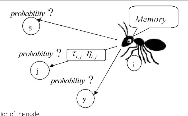
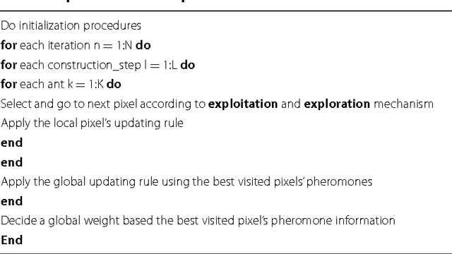
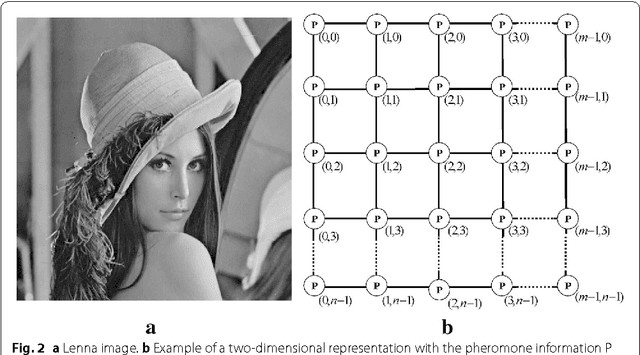
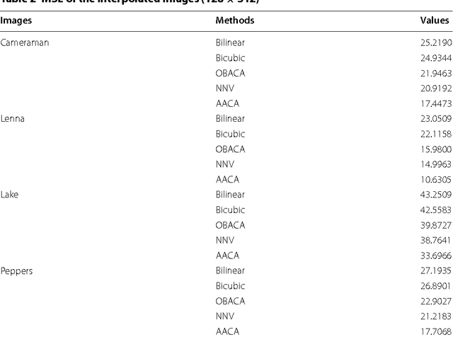
Abstract:This paper presents an advance on image interpolation based on ant colony algorithm (AACA) for high-resolution image scaling. The difference between the proposed algorithm and the previously proposed optimization of bilinear interpolation based on ant colony algorithm (OBACA) is that AACA uses global weighting, whereas OBACA uses a local weighting scheme. The strength of the proposed global weighting of the AACA algorithm depends on employing solely the pheromone matrix information present on any group of four adjacent pixels to decide which case deserves a maximum global weight value or not. Experimental results are further provided to show the higher performance of the proposed AACA algorithm with reference to the algorithms mentioned in this paper.
* 17 pages, 14 figures, 3 tables
Advanced Image Enhancement Method for Distant Vessels and Structures in Capsule Endoscopy
Apr 08, 2021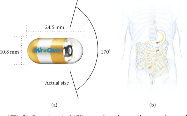
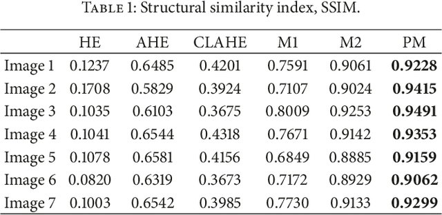
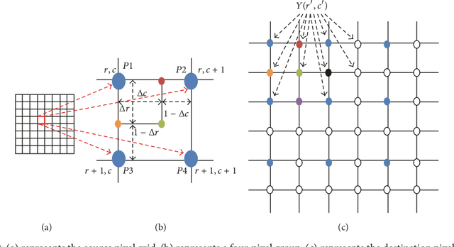
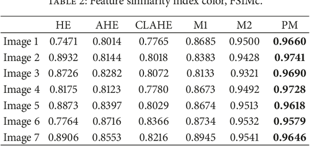
Abstract:This paper proposes an advanced method for contrast enhancement of capsule endoscopic images, with the main objective to obtain sufficient information about the vessels and structures in more distant (or darker) parts of capsule endoscopic images. The proposed method (PM) combines two algorithms for the enhancement of darker and brighter areas of capsule endoscopic images, respectively. The half-unit weighted bilinear algorithm (HWB) proposed in our previous work is used to enhance darker areas according to the darker map content of its HSV's component V. Enhancement of brighter areas is achieved thanks to the novel thresholded weighted-bilinear algorithm (TWB) developed to avoid overexposure and enlargement of specular highlight spots while preserving the hue, in such areas. The TWB performs enhancement operations following a gradual increment of the brightness of the brighter map content of its HSV's component V. In other words, the TWB decreases its averaged-weights as the intensity content of the component V increases. Extensive experimental demonstrations were conducted, and based on evaluation of the reference and PM enhanced images, a gastroenterologist ({\O}H) concluded that the PM enhanced images were the best ones based on the information about the vessels, contrast in the images, and the view or visibility of the structures in more distant parts of the capsule endoscopy images.
* 8 pages, 12 figures, 4 tables
Challenges of 3D Surface Reconstruction in Capsule Endoscopy
Mar 18, 2021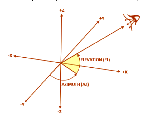
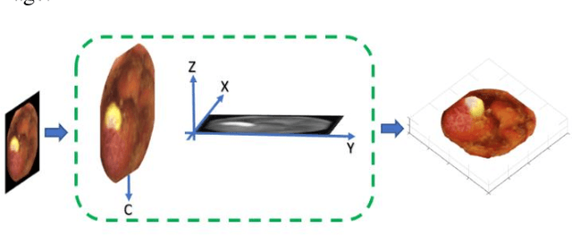
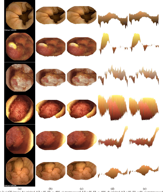
Abstract:There are currently many challenges related to three-dimensional (3D) surface reconstruction using capsule endoscopy (CE) images. There are also challenges related to viewing the content of reconstructed 3D surfaces. In this preliminary investigation, the author focuses on the latter and evaluates their effects on the content of reconstructed 3D surfaces using CE images. The evaluation of such challenges is preliminarily conducted into two parts. The first part focuses on the comparison of the content of 3D surfaces reconstructed using both preprocessed and non-preprocessed CE images. The second part focuses on the comparison of the content of 3D surfaces viewed at the same azimuth angles and different elevation angles of the line-of-sight. The experiments demonstrated the need for generalizable line-of-sight and advanced CE image preprocessing means as well as further research in 3D surface reconstruction.
Effects of Image Size on Deep Learning
Jan 27, 2021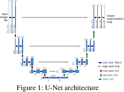
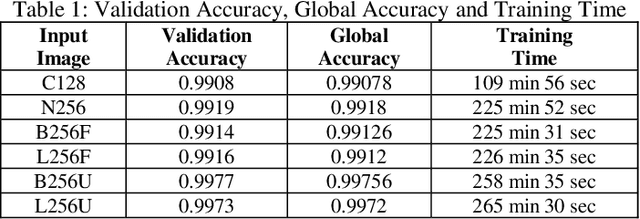
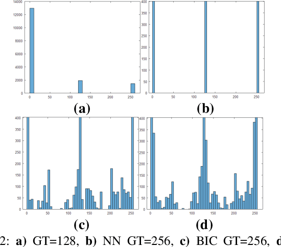

Abstract:The question is: what size of the region of interest is likely to lead to better training outcomes? To answer this: The U-net is used for semantic segmentation. Image interpolation algorithms are used to double the cropped image size and create new datasets. Depending on the selected image interpolation algorithm category, non-original classes are created in the ground truth images thus a filtering strategy is introduced to remove such spurious classes. Evaluation results of effects on the myocardium segmentation and quantification of the myocardial infarction are provided and discussed.
Evaluation of deep learning-based myocardial infarction quantification using Segment CMR software
Dec 16, 2020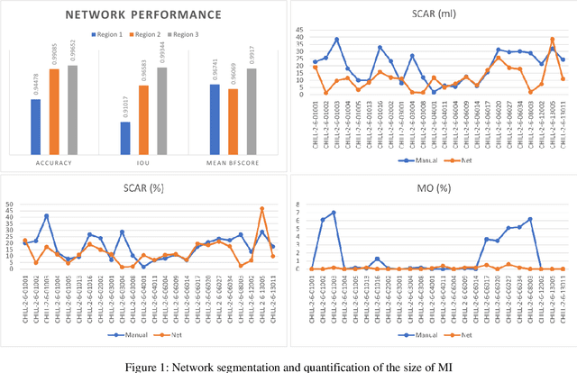
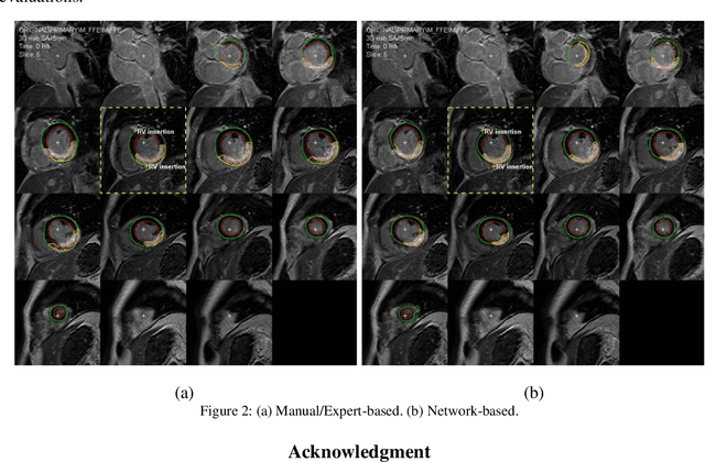
Abstract:In this paper, the author evaluates the preliminary work related to automating the quantification of the size of the myocardial infarction (MI) using deep learning in Segment cardiovascular magnetic resonance (CMR) software. Here, deep learning is used to automate the segmentation of myocardial boundaries before triggering the automatic quantification of the size of the MI using the expectation-maximization, weighted intensity, a priori information (EWA) algorithm incorporated in the Segment CMR software. Experimental evaluation of the size of the MI shows that more than 50 % (average infarct scar volume), 75% (average infarct scar percentage), and 65 % (average microvascular obstruction percentage) of the network-based results are approximately very close to the expert delineation-based results. Also, in an experiment involving the visualization of myocardial and infarct contours, in all images of the selected stack, the network and expert-based results tie in terms of the number of infarcted and contoured images.
Effect of the regularization hyperparameter on deep learning-based segmentation in LGE-MRI
Dec 15, 2020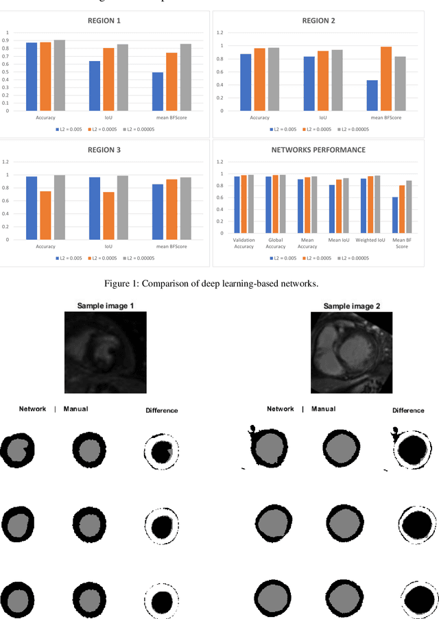
Abstract:In this work, the author aims at demonstrating the extent to which the arbitrary selection of the L2 regularization hyperparameter can affect the outcome of deep learning-based segmentation in LGE-MRI. Here, arbitrary L2 regularization values are used to create different deep learning-based segmentation networks. Also, the author adopts the manual adjustment or tunning, of other deep learning hyperparameters, to be done only when 10% of all epochs are reached before achieving the 90% validation accuracy. The experimental comparisons demonstrate that small L2 regularization values can lead to better segmentation of the myocardial boundaries.
 Add to Chrome
 Add to Firefox
 Add to Edge