Øistein Hovde
Evaluating clinical diversity and plausibility of synthetic capsule endoscopic images
Jan 16, 2023Abstract:Wireless Capsule Endoscopy (WCE) is being increasingly used as an alternative imaging modality for complete and non-invasive screening of the gastrointestinal tract. Although this is advantageous in reducing unnecessary hospital admissions, it also demands that a WCE diagnostic protocol be in place so larger populations can be effectively screened. This calls for training and education protocols attuned specifically to this modality. Like training in other modalities such as traditional endoscopy, CT, MRI, etc., a WCE training protocol would require an atlas comprising of a large corpora of images that show vivid descriptions of pathologies and abnormalities, ideally observed over a period of time. Since such comprehensive atlases are presently lacking in WCE, in this work, we propose a deep learning method for utilizing already available studies across different institutions for the creation of a realistic WCE atlas using StyleGAN. We identify clinically relevant attributes in WCE such that synthetic images can be generated with selected attributes on cue. Beyond this, we also simulate several disease progression scenarios. The generated images are evaluated for realism and plausibility through three subjective online experiments with the participation of eight gastroenterology experts from three geographical locations and a variety of years of experience. The results from the experiments indicate that the images are highly realistic and the disease scenarios plausible. The images comprising the atlas are available publicly for use in training applications as well as supplementing real datasets for deep learning.
Learning More for Free - A Multi Task Learning Approach for Improved Pathology Classification in Capsule Endoscopy
Jun 30, 2021

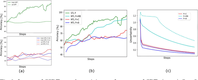

Abstract:The progress in Computer Aided Diagnosis (CADx) of Wireless Capsule Endoscopy (WCE) is thwarted by the lack of data. The inadequacy in richly representative healthy and abnormal conditions results in isolated analyses of pathologies, that can not handle realistic multi-pathology scenarios. In this work, we explore how to learn more for free, from limited data through solving a WCE multicentric, multi-pathology classification problem. Learning more implies to learning more than full supervision would allow with the same data. This is done by combining self supervision with full supervision, under multi task learning. Additionally, we draw inspiration from the Human Visual System (HVS) in designing self supervision tasks and investigate if seemingly ineffectual signals within the data itself can be exploited to gain performance, if so, which signals would be better than others. Further, we present our analysis of the high level features as a stepping stone towards more robust multi-pathology CADx in WCE.
Advanced Image Enhancement Method for Distant Vessels and Structures in Capsule Endoscopy
Apr 08, 2021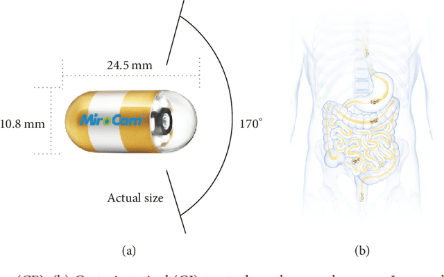
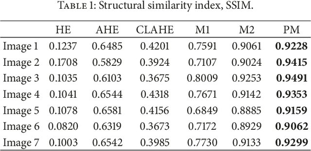
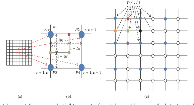
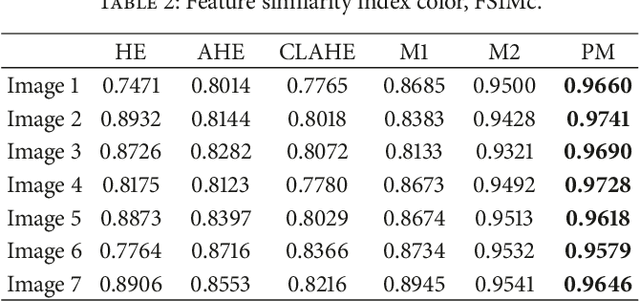
Abstract:This paper proposes an advanced method for contrast enhancement of capsule endoscopic images, with the main objective to obtain sufficient information about the vessels and structures in more distant (or darker) parts of capsule endoscopic images. The proposed method (PM) combines two algorithms for the enhancement of darker and brighter areas of capsule endoscopic images, respectively. The half-unit weighted bilinear algorithm (HWB) proposed in our previous work is used to enhance darker areas according to the darker map content of its HSV's component V. Enhancement of brighter areas is achieved thanks to the novel thresholded weighted-bilinear algorithm (TWB) developed to avoid overexposure and enlargement of specular highlight spots while preserving the hue, in such areas. The TWB performs enhancement operations following a gradual increment of the brightness of the brighter map content of its HSV's component V. In other words, the TWB decreases its averaged-weights as the intensity content of the component V increases. Extensive experimental demonstrations were conducted, and based on evaluation of the reference and PM enhanced images, a gastroenterologist ({\O}H) concluded that the PM enhanced images were the best ones based on the information about the vessels, contrast in the images, and the view or visibility of the structures in more distant parts of the capsule endoscopy images.
* 8 pages, 12 figures, 4 tables
Y-Net: A deep Convolutional Neural Network for Polyp Detection
Jun 05, 2018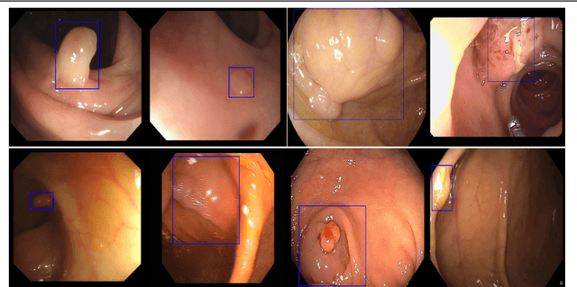

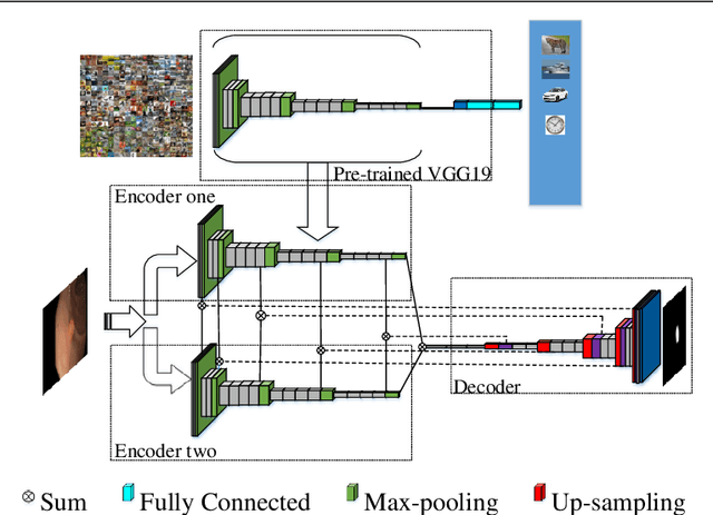

Abstract:Colorectal polyps are important precursors to colon cancer, the third most common cause of cancer mortality for both men and women. It is a disease where early detection is of crucial importance. Colonoscopy is commonly used for early detection of cancer and precancerous pathology. It is a demanding procedure requiring significant amount of time from specialized physicians and nurses, in addition to a significant miss-rates of polyps by specialists. Automated polyp detection in colonoscopy videos has been demonstrated to be a promising way to handle this problem. {However, polyps detection is a challenging problem due to the availability of limited amount of training data and large appearance variations of polyps. To handle this problem, we propose a novel deep learning method Y-Net that consists of two encoder networks with a decoder network. Our proposed Y-Net method} relies on efficient use of pre-trained and un-trained models with novel sum-skip-concatenation operations. Each of the encoders are trained with encoder specific learning rate along the decoder. Compared with the previous methods employing hand-crafted features or 2-D/3-D convolutional neural network, our approach outperforms state-of-the-art methods for polyp detection with 7.3% F1-score and 13% recall improvement.
 Add to Chrome
 Add to Firefox
 Add to Edge