Andrea Behanova
Convolutional Neural Networks for Automatic Detection of Intact Adenovirus from TEM Imaging with Debris, Broken and Artefacts Particles
Nov 06, 2023


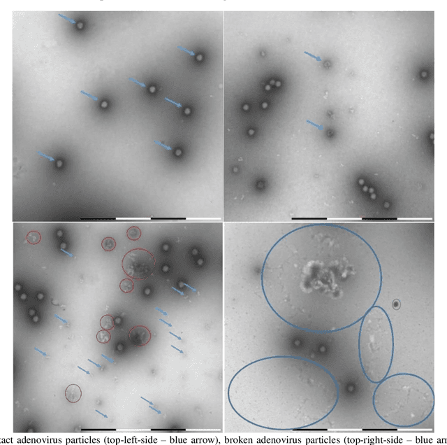
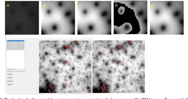
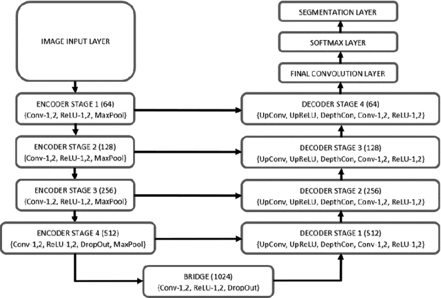
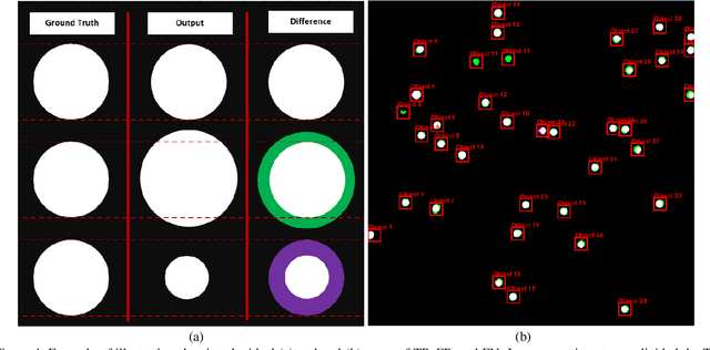
Abstract:Regular monitoring of the primary particles and purity profiles of a drug product during development and manufacturing processes is essential for manufacturers to avoid product variability and contamination. Transmission electron microscopy (TEM) imaging helps manufacturers predict how changes affect particle characteristics and purity for virus-based gene therapy vector products and intermediates. Since intact particles can characterize efficacious products, it is beneficial to automate the detection of intact adenovirus against a non-intact-viral background mixed with debris, broken, and artefact particles. In the presence of such particles, detecting intact adenoviruses becomes more challenging. To overcome the challenge, due to such a presence, we developed a software tool for semi-automatic annotation and segmentation of adenoviruses and a software tool for automatic segmentation and detection of intact adenoviruses in TEM imaging systems. The developed semi-automatic tool exploited conventional image analysis techniques while the automatic tool was built based on convolutional neural networks and image analysis techniques. Our quantitative and qualitative evaluations showed outstanding true positive detection rates compared to false positive and negative rates where adenoviruses were nicely detected without mistaking them for real debris, broken adenoviruses, and/or staining artefacts.
gACSON software for automated segmentation and morphology analyses of myelinated axons in 3D electron microscopy
Dec 13, 2021


Abstract:Background and Objective: Advances in electron microscopy (EM) now allow three-dimensional (3D) imaging of hundreds of micrometers of tissue with nanometer-scale resolution, providing new opportunities to study the ultrastructure of the brain. In this work, we introduce a freely available gACSON software for visualization, segmentation, assessment, and morphology analysis of myelinated axons in 3D-EM volumes of brain tissue samples. Methods: The gACSON software is equipped with a graphical user interface (GUI). It automatically segments the intra-axonal space of myelinated axons and their corresponding myelin sheaths and allows manual segmentation, proofreading, and interactive correction of the segmented components. gACSON analyzes the morphology of myelinated axons, such as axonal diameter, axonal eccentricity, myelin thickness, or g-ratio. Results: We illustrate the use of gACSON by segmenting and analyzing myelinated axons in six 3D-EM volumes of rat somatosensory cortex after sham surgery or traumatic brain injury (TBI). Our results suggest that the equivalent diameter of myelinated axons in somatisensory cortex was decreased in TBI animals five months after the injury. Conclusions: Our results indicate that gACSON is a valuable tool for visualization, segmentation, assessment, and morphology analysis of myelinated axons in 3D-EM volumes. gACSON is freely available at https://github.com/AndreaBehan/g-ACSON under the MIT license.
 Add to Chrome
 Add to Firefox
 Add to Edge