Nima Emami
AI enhanced finite element multiscale modelling and structural uncertainty analysis of a functionally graded porous beam
Nov 02, 2022


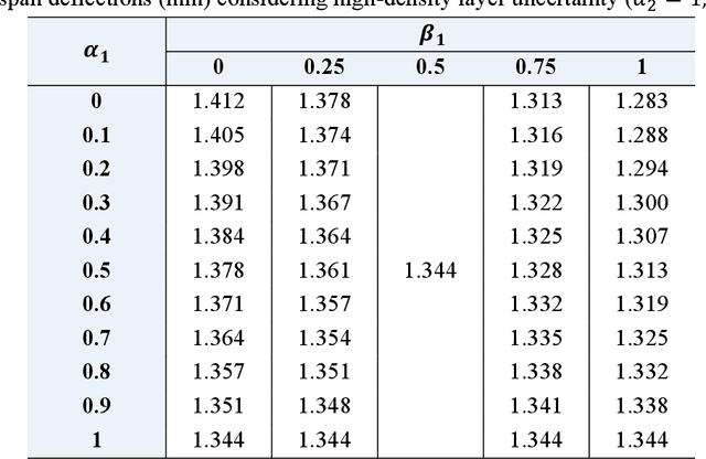
Abstract:The local geometrical randomness of metal foams brings complexities to the performance prediction of porous structures. Although the relative density is commonly deemed as the key factor, the stochasticity of internal cell sizes and shapes has an apparent effect on the porous structural behaviour but the corresponding measurement is challenging. To address this issue, we are aimed to develop an assessment strategy for efficiently examining the foam properties by combining multiscale modelling and deep learning. The multiscale modelling is based on the finite element (FE) simulation employing representative volume elements (RVEs) with random cellular morphologies, mimicking the typical features of closed-cell Aluminium foams. A deep learning database is constructed for training the designed convolutional neural networks (CNNs) to establish a direct link between the mesoscopic porosity characteristics and the effective Youngs modulus of foams. The error range of CNN models leads to an uncertain mechanical performance, which is further evaluated in a structural uncertainty analysis on the FG porous three-layer beam consisting of two thin high-density layers and a thick low-density one, where the imprecise CNN predicted moduli are represented as triangular fuzzy numbers in double parametric form. The uncertain beam bending deflections under a mid-span point load are calculated with the aid of Timoshenko beam theory and the Ritz method. Our findings suggest the success in training CNN models to estimate RVE modulus using images with an average error of 5.92%. The evaluation of FG porous structures can be significantly simplified with the proposed method and connects to the mesoscopic cellular morphologies without establishing the mechanics model for local foams.
A deep learned nanowire segmentation model using synthetic data augmentation
Sep 28, 2021


Abstract:Automatized object identification and feature analysis of experimental image data are indispensable for data-driven material science; deep-learning-based segmentation algorithms have been shown to be a promising technique to achieve this goal. However, acquiring high-resolution experimental images and assigning labels in order to train such algorithms is challenging and costly in terms of both time and labor. In the present work, we apply synthetic images, which resemble the experimental image data in terms of geometrical and visual features, to train state-of-art deep learning-based Mask R-CNN algorithms to segment vanadium pentoxide (V2O5) nanowires, a canonical cathode material, within optical intensity-based images from spectromicroscopy. The performance evaluation demonstrates that even though the deep learning model is trained on pure synthetically generated structures, it can segment real optical intensity-based spectromicroscopy images of complex V2O5 nanowire structures in overlapped particle networks, thus providing reliable statistical information. The model can further be used to segment nanowires in scanning electron microscopy (SEM) images, which are fundamentally different from the training dataset known to the model. The proposed methodology of using a purely synthetic dataset to train the deep learning model can be extended to any optical intensity-based images of variable particle morphology, extent of agglomeration, material class, and beyond.
 Add to Chrome
 Add to Firefox
 Add to Edge