Miguel Angel Gonzalez Ballester
Decoder-Free Supervoxel GNN for Accurate Brain-Tumor Localization in Multi-Modal MRI
Jan 20, 2026Abstract:Modern vision backbones for 3D medical imaging typically process dense voxel grids through parameter-heavy encoder-decoder structures, a design that allocates a significant portion of its parameters to spatial reconstruction rather than feature learning. Our approach introduces SVGFormer, a decoder-free pipeline built upon a content-aware grouping stage that partitions the volume into a semantic graph of supervoxels. Its hierarchical encoder learns rich node representations by combining a patch-level Transformer with a supervoxel-level Graph Attention Network, jointly modeling fine-grained intra-region features and broader inter-regional dependencies. This design concentrates all learnable capacity on feature encoding and provides inherent, dual-scale explainability from the patch to the region level. To validate the framework's flexibility, we trained two specialized models on the BraTS dataset: one for node-level classification and one for tumor proportion regression. Both models achieved strong performance, with the classification model achieving a F1-score of 0.875 and the regression model a MAE of 0.028, confirming the encoder's ability to learn discriminative and localized features. Our results establish that a graph-based, encoder-only paradigm offers an accurate and inherently interpretable alternative for 3D medical image representation.
Fetpype: An Open-Source Pipeline for Reproducible Fetal Brain MRI Analysis
Dec 19, 2025Abstract:Fetal brain Magnetic Resonance Imaging (MRI) is crucial for assessing neurodevelopment in utero. However, analyzing this data presents significant challenges due to fetal motion, low signal-to-noise ratio, and the need for complex multi-step processing, including motion correction, super-resolution reconstruction, segmentation, and surface extraction. While various specialized tools exist for individual steps, integrating them into robust, reproducible, and user-friendly workflows that go from raw images to processed volumes is not straightforward. This lack of standardization hinders reproducibility across studies and limits the adoption of advanced analysis techniques for researchers and clinicians. To address these challenges, we introduce Fetpype, an open-source Python library designed to streamline and standardize the preprocessing and analysis of T2-weighted fetal brain MRI data. Fetpype is publicly available on GitHub at https://github.com/fetpype/fetpype.
Attri-VAE: attribute-based, disentangled and interpretable representations of medical images with variational autoencoders
Mar 27, 2022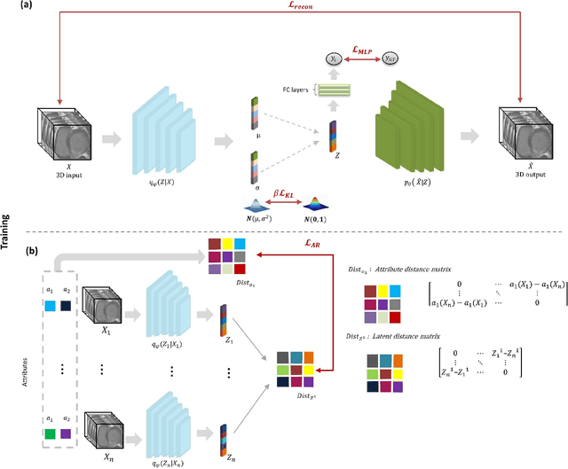
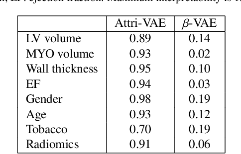
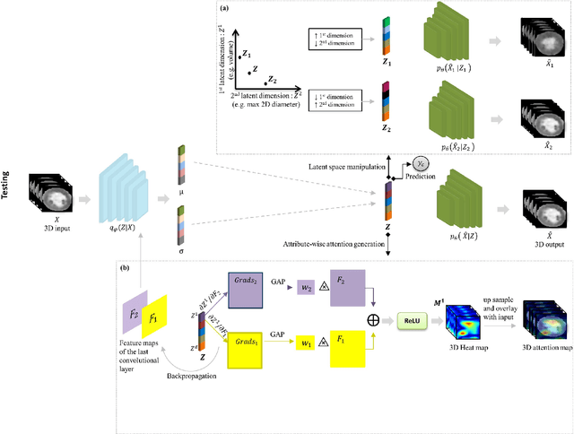

Abstract:Deep learning (DL) methods where interpretability is intrinsically considered as part of the model are required to better understand the relationship of clinical and imaging-based attributes with DL outcomes, thus facilitating their use in reasoning medical decisions. Latent space representations built with variational autoencoders (VAE) do not ensure individual control of data attributes. Attribute-based methods enforcing attribute disentanglement have been proposed in the literature for classical computer vision tasks in benchmark data. In this paper, we propose a VAE approach, the Attri-VAE, that includes an attribute regularization term to associate clinical and medical imaging attributes with different regularized dimensions in the generated latent space, enabling a better disentangled interpretation of the attributes. Furthermore, the generated attention maps explained the attribute encoding in the regularized latent space dimensions. The Attri-VAE approach analyzed healthy and myocardial infarction patients with clinical, cardiac morphology, and radiomics attributes. The proposed model provided an excellent trade-off between reconstruction fidelity, disentanglement, and interpretability, outperforming state-of-the-art VAE approaches according to several quantitative metrics. The resulting latent space allowed the generation of realistic synthetic data in the trajectory between two distinct input samples or along a specific attribute dimension to better interpret changes between different cardiac conditions.
A radiomics approach to analyze cardiac alterations in hypertension
Jul 21, 2020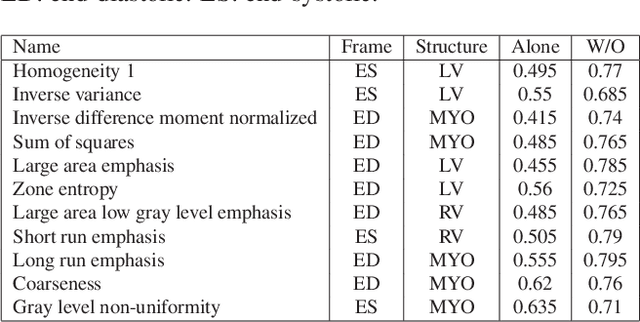
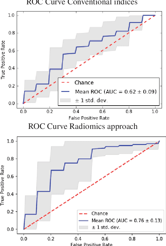
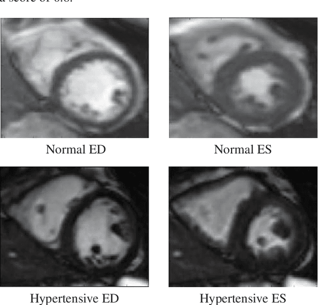
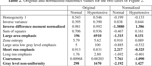
Abstract:Hypertension is a medical condition that is well-established as a risk factor for many major diseases. For example, it can cause alterations in the cardiac structure and function over time that can lead to heart related morbidity and mortality. However, at the subclinical stage, these changes are subtle and cannot be easily captured using conventional cardiovascular indices calculated from clinical cardiac imaging. In this paper, we describe a radiomics approach for identifying intermediate imaging phenotypes associated with hypertension. The method combines feature selection and machine learning techniques to identify the most subtle as well as complex structural and tissue changes in hypertensive subgroups as compared to healthy individuals. Validation based on a sample of asymptomatic hearts that include both hypertensive and non-hypertensive cases demonstrate that the proposed radiomics model is capable of detecting intensity and textural changes well beyond the capabilities of conventional imaging phenotypes, indicating its potential for improved understanding of the longitudinal effects of hypertension on cardiovascular health and disease.
Computational Anatomy for Multi-Organ Analysis in Medical Imaging: A Review
Dec 20, 2018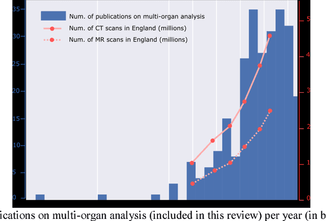
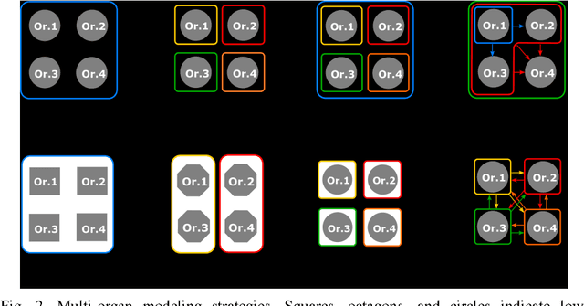
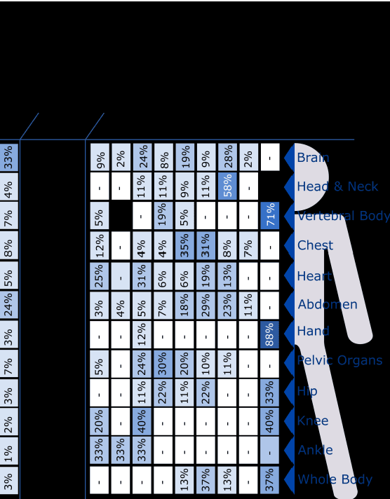
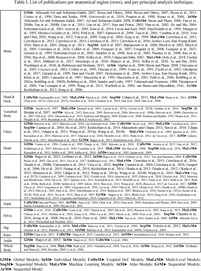
Abstract:The medical image analysis field has traditionally been focused on the development of organ-, and disease-specific methods. Recently, the interest in the development of more 20 comprehensive computational anatomical models has grown, leading to the creation of multi-organ models. Multi-organ approaches, unlike traditional organ-specific strategies, incorporate inter-organ relations into the model, thus leading to a more accurate representation of the complex human anatomy. Inter-organ relations are not only spatial, but also functional and physiological. Over the years, the strategies 25 proposed to efficiently model multi-organ structures have evolved from the simple global modeling, to more sophisticated approaches such as sequential, hierarchical, or machine learning-based models. In this paper, we present a review of the state of the art on multi-organ analysis and associated computation anatomy methodology. The manuscript follows a methodology-based classification of the different techniques 30 available for the analysis of multi-organs and multi-anatomical structures, from techniques using point distribution models to the most recent deep learning-based approaches. With more than 300 papers included in this review, we reflect on the trends and challenges of the field of computational anatomy, the particularities of each anatomical region, and the potential of multi-organ analysis to increase the impact of 35 medical imaging applications on the future of healthcare.
 Add to Chrome
 Add to Firefox
 Add to Edge