Maciej Pech
Predicting 4D Liver MRI for MR-guided Interventions
Feb 25, 2022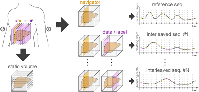


Abstract:Organ motion poses an unresolved challenge in image-guided interventions. In the pursuit of solving this problem, the research field of time-resolved volumetric magnetic resonance imaging (4D MRI) has evolved. However, current techniques are unsuitable for most interventional settings because they lack sufficient temporal and/or spatial resolution or have long acquisition times. In this work, we propose a novel approach for real-time, high-resolution 4D MRI with large fields of view for MR-guided interventions. To this end, we trained a convolutional neural network (CNN) end-to-end to predict a 3D liver MRI that correctly predicts the liver's respiratory state from a live 2D navigator MRI of a subject. Our method can be used in two ways: First, it can reconstruct near real-time 4D MRI with high quality and high resolution (209x128x128 matrix size with isotropic 1.8mm voxel size and 0.6s/volume) given a dynamic interventional 2D navigator slice for guidance during an intervention. Second, it can be used for retrospective 4D reconstruction with a temporal resolution of below 0.2s/volume for motion analysis and use in radiation therapy. We report a mean target registration error (TRE) of 1.19 $\pm$0.74mm, which is below voxel size. We compare our results with a state-of-the-art retrospective 4D MRI reconstruction. Visual evaluation shows comparable quality. We show that small training sizes with short acquisition times down to 2min can already achieve promising results and 24min are sufficient for high quality results. Because our method can be readily combined with earlier methods, acquisition time can be further decreased while also limiting quality loss. We show that an end-to-end, deep learning formulation is highly promising for 4D MRI reconstruction.
Joint Liver and Hepatic Lesion Segmentation using a Hybrid CNN with Transformer Layers
Jan 26, 2022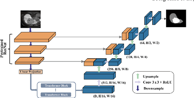


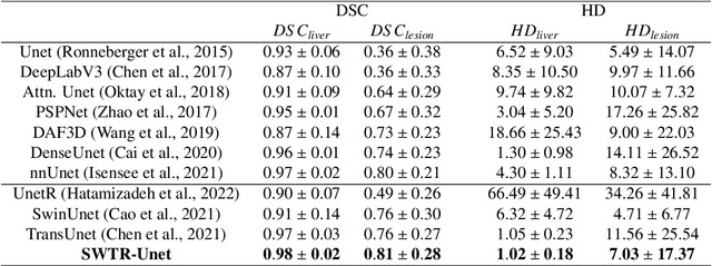
Abstract:Deep learning-based segmentation of the liver and hepatic lesions therein steadily gains relevance in clinical practice due to the increasing incidence of liver cancer each year. Whereas various network variants with overall promising results in the field of medical image segmentation have been developed over the last years, almost all of them struggle with the challenge of accurately segmenting hepatic lesions. This lead to the idea of combining elements of convolutional and transformerbased architectures to overcome the existing limitations. This work presents a hybrid network called SWTR-Unet, consisting of a pretrained ResNet, transformer blocks as well as a common Unet-style decoder path. This network was applied to clinical liver MRI, as well as to the publicly available CT data of the liver tumor segmentation (LiTS) challenge. Additionally, multiple state-of-the-art networks were implemented and applied to both datasets, ensuring a direct comparability. Furthermore, correlation analysis and an ablation study were carried out, to investigate various influencing factors on the segmentation accuracy of our presented method. With Dice similarity scores of averaged 98 +- 2 % for liver and 81 +- 28 % lesion segmentation on the MRI dataset and 97 +- 2 % and 79 +- 25 %, respectively on the CT dataset, the proposed SWTR-Unet outperforms each of the additionally implemented state-of-the-art networks. The achieved segmentation accuracy was found to be on par with manually performed expert segmentations as indicated by interobserver variabilities for liver lesion segmentation. In conclusion, the presented method could save valuable time and resources in clinical practice.
4D MRI: Robust sorting of free breathing MRI slices for use in interventional settings
Oct 04, 2019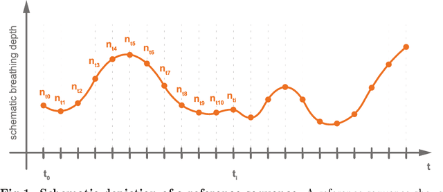
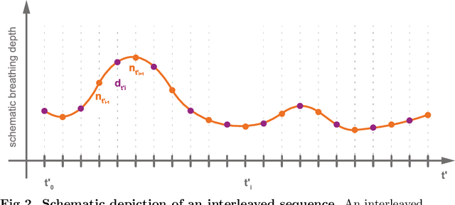
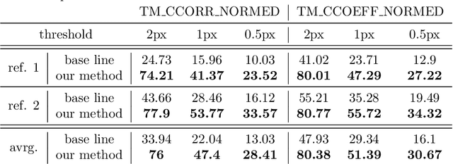
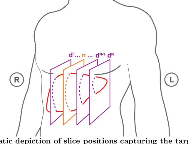
Abstract:Purpose: We aim to develop a robust 4D MRI method for large FOVs enabling the extraction of irregular respiratory motion that is readily usable with all MRI machines and thus applicable to support a wide range of interventional settings. Method: We propose a 4D MRI reconstruction method to capture an arbitrary number of breathing states. It uses template updates in navigator slices and search regions for fast and robust vessel cross-section tracking. It captures FOVs of 255 mm x 320 mm x 228 mm at a spatial resolution of 1.82 mm x 1.82 mm x 4mm and temporal resolution of 200ms. To validate the method, a total of 38 4D MRIs of 13 healthy subjects were reconstructed. A quantitative evaluation of the reconstruction rate and speed of both the new and baseline method was performed. Additionally, a study with ten radiologists was conducted to assess the subjective reconstruction quality of both methods. Results: Our results indicate improved mean reconstruction rates compared to the baseline method (79.4\% vs. 45.5\%) and improved mean reconstruction times (24s vs. 73s) per subject. Interventional radiologists perceive the reconstruction quality of our method as higher compared to the baseline (262.5 points vs. 217.5 points, p=0.02). Conclusions: Template updates are an effective and efficient way to increase 4D MRI reconstruction rates and to achieve better reconstruction quality. Search regions reduce reconstruction time. These improvements increase the applicability of 4D MRI as base for seamless support of interventional image guidance in percutaneous interventions.
 Add to Chrome
 Add to Firefox
 Add to Edge