Karl F. Böhringer
Large FOV short-wave infrared meta-lens for scanning fiber endoscopy
Dec 21, 2022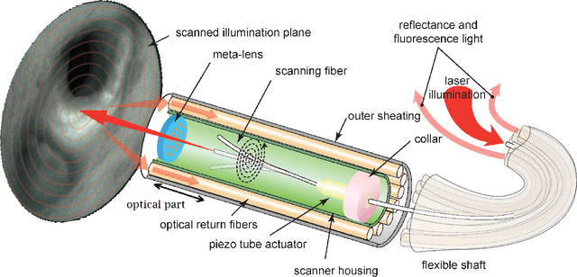
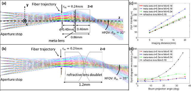
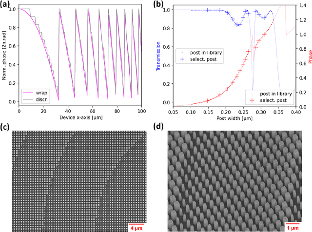
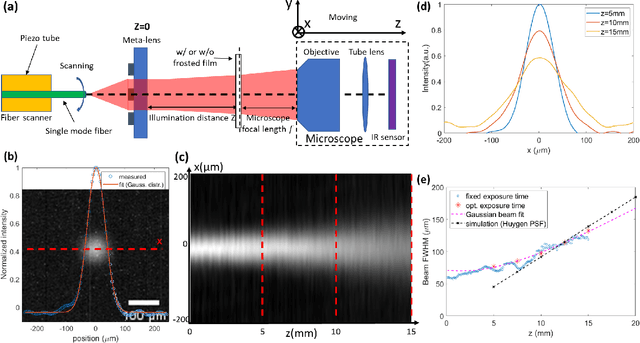
Abstract:The scanning fiber endoscope (SFE), an ultra-small optical imaging device with a large field-of-view (FOV) for having a clear forward view into the interior of blood vessels, has great potential in the cardio-vascular disease diagnosis and surgery assistance, which is one of the key applications for short-wave infrared (SWIR) biomedical imaging. The state-of-the-art SFE system uses a miniaturized refractive spherical lens doublet for beam projection. A meta-lens is a promising alternative which can be made much thinner and has fewer off-axis aberrations than its refractive counterpart. We report an SFE system with meta-lens working at 1310nm to achieve a resolution ($\sim 140\mu m$ at the center of field and the imaging distance of $15mm$), FOV ($\sim 70 \circ$), and depth-of-focus (DOF $\sim 15mm$), which are comparable to a state-of-the-art refractive lens SFE. The use of the meta-lens reduces the length of the optical track from $1.2mm$ to $0.86mm$. The resolution of our meta-lens based SFE drops by less than a factor of $2$ at the edge of the FOV, while the refractive lens counterpart has a $\sim 3$ times resolution degradation. These results show the promise of integrating a meta-lens into an endoscope for device minimization and optical performance improvement.
Foveated Thermal Computational Imaging in the Wild Using All-Silicon Meta-Optics
Dec 13, 2022Abstract:Foveated imaging provides a better tradeoff between situational awareness (field of view) and resolution and is critical in long-wavelength infrared regimes because of the size, weight, power, and cost of thermal sensors. We demonstrate computational foveated imaging by exploiting the ability of a meta-optical frontend to discriminate between different polarization states and a computational backend to reconstruct the captured image/video. The frontend is a three-element optic: the first element which we call the "foveal" element is a metalens that focuses s-polarized light at a distance of $f_1$ without affecting the p-polarized light; the second element which we call the "perifoveal" element is another metalens that focuses p-polarized light at a distance of $f_2$ without affecting the s-polarized light. The third element is a freely rotating polarizer that dynamically changes the mixing ratios between the two polarization states. Both the foveal element (focal length = 150mm; diameter = 75mm), and the perifoveal element (focal length = 25mm; diameter = 25mm) were fabricated as polarization-sensitive, all-silicon, meta surfaces resulting in a large-aperture, 1:6 foveal expansion, thermal imaging capability. A computational backend then utilizes a deep image prior to separate the resultant multiplexed image or video into a foveated image consisting of a high-resolution center and a lower-resolution large field of view context. We build a first-of-its-kind prototype system and demonstrate 12 frames per second real-time, thermal, foveated image, and video capture in the wild.
Inverse-Designed Meta-Optics with Spectral-Spatial Engineered Response to Mimic Color Perception
Apr 28, 2022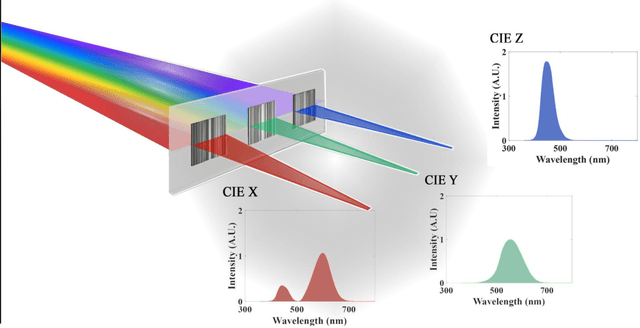

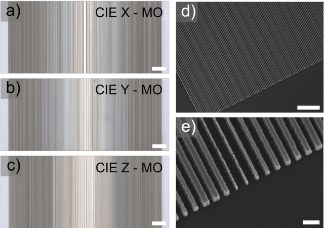
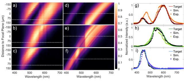
Abstract:Meta-optics have rapidly become a major research field within the optics and photonics community, strongly driven by the seemingly limitless opportunities made possible by controlling optical wavefronts through interaction with arrays of sub-wavelength scatterers. As more and more modalities are explored, the design strategies to achieve desired functionalities become increasingly demanding, necessitating more advanced design techniques. Herein, the inverse-design approach is utilized to create a set of single-layer meta-optics that simultaneously focus light and shape the spectra of focused light without using any filters. Thus, both spatial and spectral properties of the meta-optics are optimized, resulting in spectra that mimic the color matching functions of the CIE 1931 XYZ color space, which links the distributions of wavelengths in light and the color perception of a human eye. Experimental demonstrations of these meta-optics show qualitative agreement with the theoretical predictions and help elucidate the focusing mechanism of these devices.
 Add to Chrome
 Add to Firefox
 Add to Edge