John S Zelek
PitcherNet: Powering the Moneyball Evolution in Baseball Video Analytics
May 13, 2024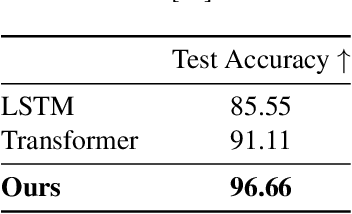
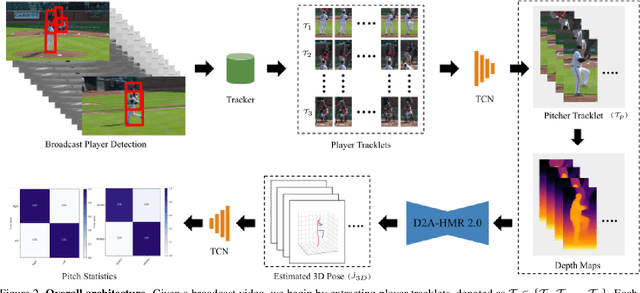
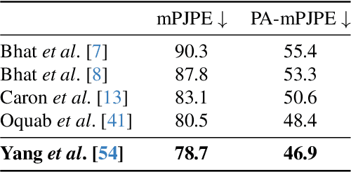
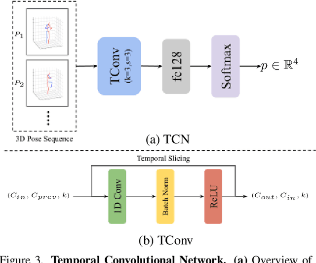
Abstract:In the high-stakes world of baseball, every nuance of a pitcher's mechanics holds the key to maximizing performance and minimizing runs. Traditional analysis methods often rely on pre-recorded offline numerical data, hindering their application in the dynamic environment of live games. Broadcast video analysis, while seemingly ideal, faces significant challenges due to factors like motion blur and low resolution. To address these challenges, we introduce PitcherNet, an end-to-end automated system that analyzes pitcher kinematics directly from live broadcast video, thereby extracting valuable pitch statistics including velocity, release point, pitch position, and release extension. This system leverages three key components: (1) Player tracking and identification by decoupling actions from player kinematics; (2) Distribution and depth-aware 3D human modeling; and (3) Kinematic-driven pitch statistics. Experimental validation demonstrates that PitcherNet achieves robust analysis results with 96.82% accuracy in pitcher tracklet identification, reduced joint position error by 1.8mm and superior analytics compared to baseline methods. By enabling performance-critical kinematic analysis from broadcast video, PitcherNet paves the way for the future of baseball analytics by optimizing pitching strategies, preventing injuries, and unlocking a deeper understanding of pitcher mechanics, forever transforming the game.
PixelBNN: Augmenting the PixelCNN with batch normalization and the presentation of a fast architecture for retinal vessel segmentation
Dec 19, 2017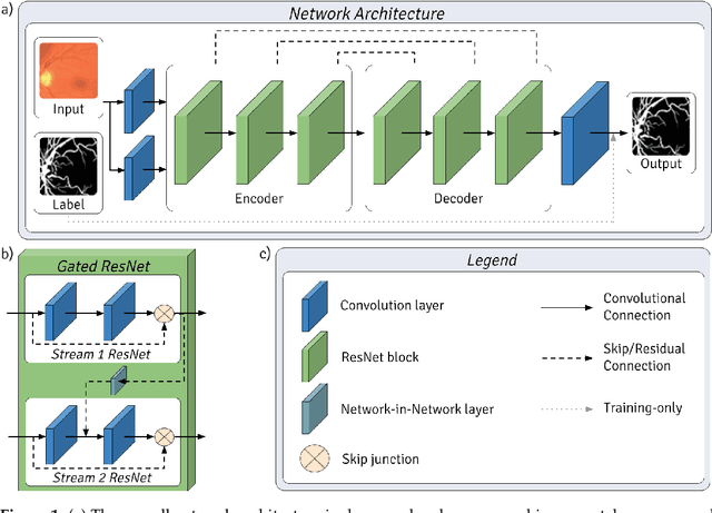
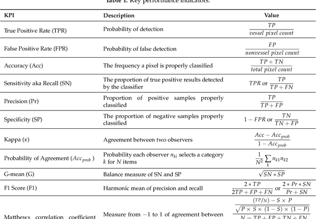
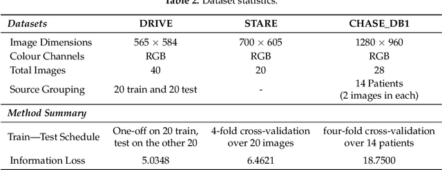
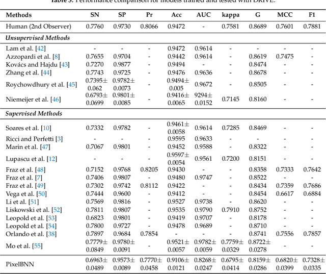
Abstract:Analysis of retinal fundus images is essential for eye-care physicians in the diagnosis, care and treatment of patients. Accurate fundus and/or retinal vessel maps give rise to longitudinal studies able to utilize multimedia image registration and disease/condition status measurements, as well as applications in surgery preparation and biometrics. The segmentation of retinal morphology has numerous applications in assessing ophthalmologic and cardiovascular disease pathologies. The early detection of many such conditions is often the most effective method for reducing patient risk. Computer aided segmentation of the vasculature has proven to be a challenge, mainly due to inconsistencies such as noise and variations in hue and brightness that can greatly reduce the quality of fundus images. This paper presents PixelBNN, a highly efficient deep method for automating the segmentation of fundus morphologies. The model was trained, tested and cross tested on the DRIVE, STARE and CHASE\_DB1 retinal vessel segmentation datasets. Performance was evaluated using G-mean, Mathews Correlation Coefficient and F1-score. The network was 8.5 times faster than the current state-of-the-art at test time and performed comparatively well, considering a 5 to 19 times reduction in information from resizing images during preprocessing.
 Add to Chrome
 Add to Firefox
 Add to Edge