John A. Bogovic
HHMI Janelia Research Campus
Decomposing heterogeneous dynamical systems with graph neural networks
Jul 27, 2024Abstract:Natural physical, chemical, and biological dynamical systems are often complex, with heterogeneous components interacting in diverse ways. We show that graph neural networks can be designed to jointly learn the interaction rules and the structure of the heterogeneity from data alone. The learned latent structure and dynamics can be used to virtually decompose the complex system which is necessary to parameterize and infer the underlying governing equations. We tested the approach with simulation experiments of moving particles and vector fields that interact with each other. While our current aim is to better understand and validate the approach with simulated data, we anticipate it to become a generally applicable tool to uncover the governing rules underlying complex dynamics observed in nature.
Deep Learning for Isotropic Super-Resolution from Non-Isotropic 3D Electron Microscopy
Jun 09, 2017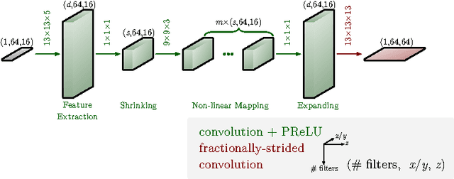

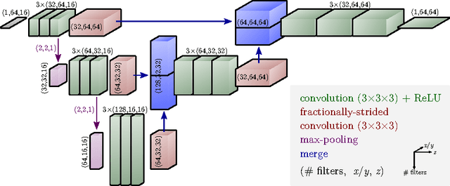

Abstract:The most sophisticated existing methods to generate 3D isotropic super-resolution (SR) from non-isotropic electron microscopy (EM) are based on learned dictionaries. Unfortunately, none of the existing methods generate practically satisfying results. For 2D natural images, recently developed super-resolution methods that use deep learning have been shown to significantly outperform the previous state of the art. We have adapted one of the most successful architectures (FSRCNN) for 3D super-resolution, and compared its performance to a 3D U-Net architecture that has not been used previously to generate super-resolution. We trained both architectures on artificially downscaled isotropic ground truth from focused ion beam milling scanning EM (FIB-SEM) and tested the performance for various hyperparameter settings. Our results indicate that both architectures can successfully generate 3D isotropic super-resolution from non-isotropic EM, with the U-Net performing consistently better. We propose several promising directions for practical application.
Image-Based Correction of Continuous and Discontinuous Non-Planar Axial Distortion in Serial Section Microscopy
Jun 17, 2016
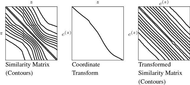
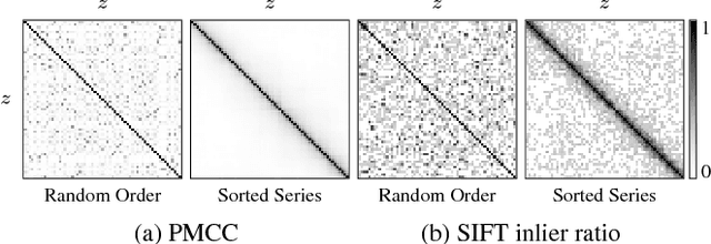
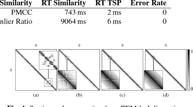
Abstract:Motivation: Serial section microscopy is an established method for detailed anatomy reconstruction of biological specimen. During the last decade, high resolution electron microscopy (EM) of serial sections has become the de-facto standard for reconstruction of neural connectivity at ever increasing scales (EM connectomics). In serial section microscopy, the axial dimension of the volume is sampled by physically removing thin sections from the embedded specimen and subsequently imaging either the block-face or the section series. This process has limited precision leading to inhomogeneous non-planar sampling of the axial dimension of the volume which, in turn, results in distorted image volumes. This includes that section series may be collected and imaged in unknown order. Results: We developed methods to identify and correct these distortions through image-based signal analysis without any additional physical apparatus or measurements. We demonstrate the efficacy of our methods in proof of principle experiments and application to real world problems. Availability and Implementation: We made our work available as libraries for the ImageJ distribution Fiji and for deployment in a high performance parallel computing environment. Our sources are open and available at http://github.com/saalfeldla/section-sort, http://github.com/saalfeldlab/em-thickness-estimation, and http://github.com/saalfeldlab/z-spacing-spark. Contact: saalfelds@janelia.hhmi.org
Robust Registration of Calcium Images by Learned Contrast Synthesis
Nov 03, 2015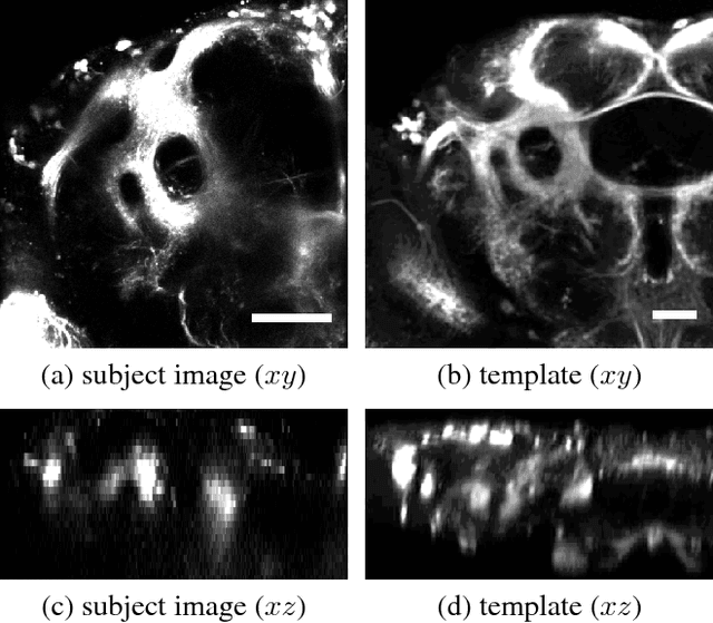
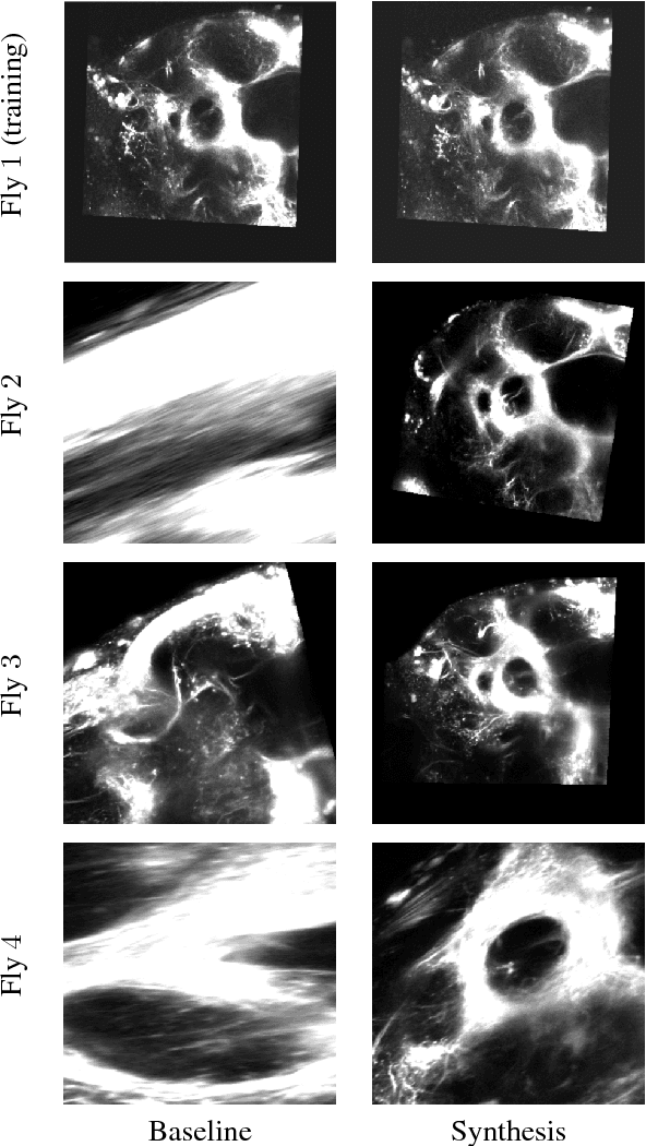

Abstract:Multi-modal image registration is a challenging task that is vital to fuse complementary signals for subsequent analyses. Despite much research into cost functions addressing this challenge, there exist cases in which these are ineffective. In this work, we show that (1) this is true for the registration of in-vivo Drosophila brain volumes visualizing genetically encoded calcium indicators to an nc82 atlas and (2) that machine learning based contrast synthesis can yield improvements. More specifically, the number of subjects for which the registration outright failed was greatly reduced (from 40% to 15%) by using a synthesized image.
Post-acquisition image based compensation for thickness variation in microscopy section series
May 27, 2015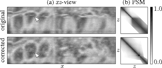
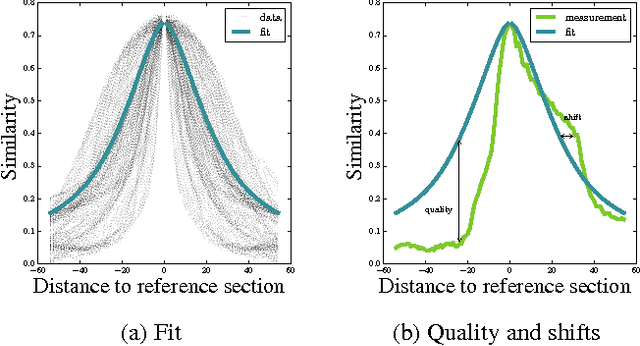
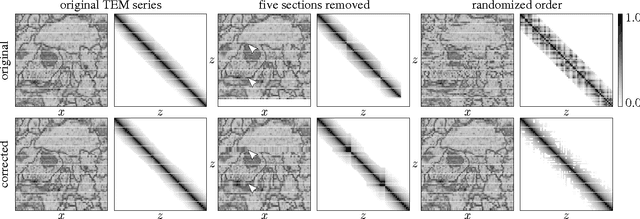
Abstract:Serial section Microscopy is an established method for volumetric anatomy reconstruction. Section series imaged with Electron Microscopy are currently vital for the reconstruction of the synaptic connectivity of entire animal brains such as that of Drosophila melanogaster. The process of removing ultrathin layers from a solid block containing the specimen, however, is a fragile procedure and has limited precision with respect to section thickness. We have developed a method to estimate the relative z-position of each individual section as a function of signal change across the section series. First experiments show promising results on both serial section Transmission Electron Microscopy (ssTEM) data and Focused Ion Beam Scanning Electron Microscopy (FIB-SEM) series. We made our solution available as Open Source plugins for the TrakEM2 software and the ImageJ distribution Fiji.
Learned versus Hand-Designed Feature Representations for 3d Agglomeration
Dec 20, 2013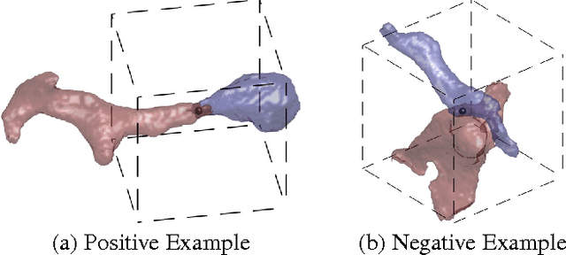
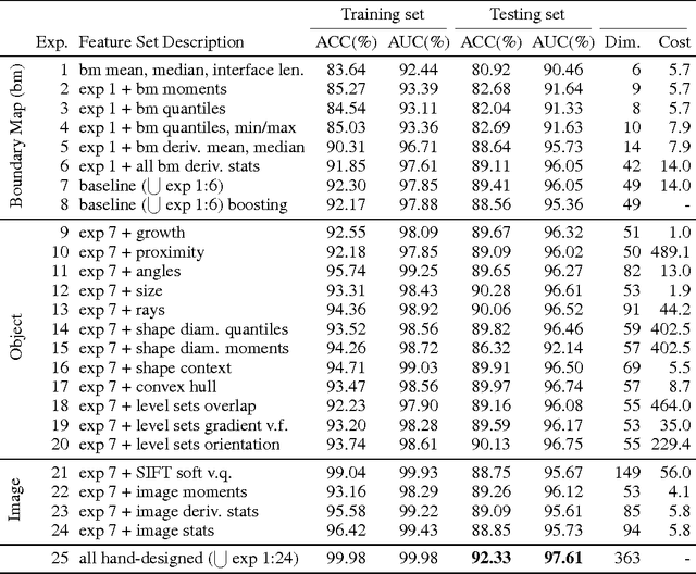

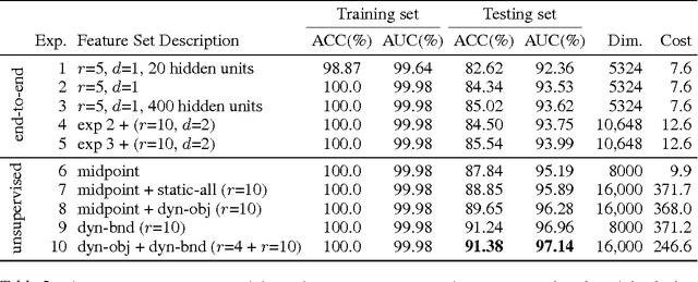
Abstract:For image recognition and labeling tasks, recent results suggest that machine learning methods that rely on manually specified feature representations may be outperformed by methods that automatically derive feature representations based on the data. Yet for problems that involve analysis of 3d objects, such as mesh segmentation, shape retrieval, or neuron fragment agglomeration, there remains a strong reliance on hand-designed feature descriptors. In this paper, we evaluate a large set of hand-designed 3d feature descriptors alongside features learned from the raw data using both end-to-end and unsupervised learning techniques, in the context of agglomeration of 3d neuron fragments. By combining unsupervised learning techniques with a novel dynamic pooling scheme, we show how pure learning-based methods are for the first time competitive with hand-designed 3d shape descriptors. We investigate data augmentation strategies for dramatically increasing the size of the training set, and show how combining both learned and hand-designed features leads to the highest accuracy.
 Add to Chrome
 Add to Firefox
 Add to Edge