Ji-Xin Cheng
Self-Supervised Elimination of Non-Independent Noise in Hyperspectral Imaging
Sep 16, 2024


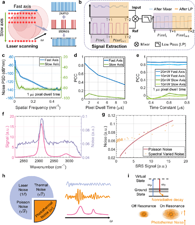
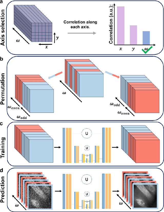
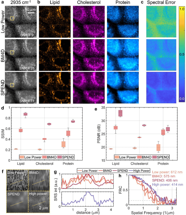
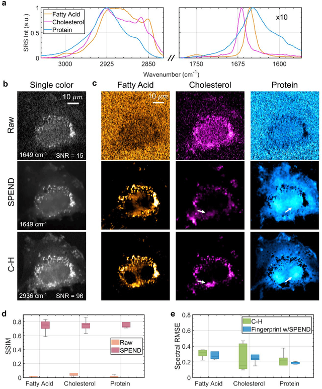
Abstract:Hyperspectral imaging has been widely used for spectral and spatial identification of target molecules, yet often contaminated by sophisticated noise. Current denoising methods generally rely on independent and identically distributed noise statistics, showing corrupted performance for non-independent noise removal. Here, we demonstrate Self-supervised PErmutation Noise2noise Denoising (SPEND), a deep learning denoising architecture tailor-made for removing non-independent noise from a single hyperspectral image stack. We utilize hyperspectral stimulated Raman scattering and mid-infrared photothermal microscopy as the testbeds, where the noise is spatially correlated and spectrally varied. Based on single hyperspectral images, SPEND permutates odd and even spectral frames to generate two stacks with identical noise properties, and uses the pairs for efficient self-supervised noise-to-noise training. SPEND achieved an 8-fold signal-to-noise improvement without having access to the ground truth data. SPEND enabled accurate mapping of low concentration biomolecules in both fingerprint and silent regions, demonstrating its robustness in sophisticated cellular environments.
High-content stimulated Raman histology of human breast cancer
Sep 20, 2023


Abstract:Histological examination is crucial for cancer diagnosis, including hematoxylin and eosin (H&E) staining for mapping morphology and immunohistochemistry (IHC) staining for revealing chemical information. Recently developed two-color stimulated Raman histology could bypass the complex tissue processing to mimic H&E-like morphology. Yet, the underlying chemical features are not revealed, compromising the effectiveness of prognostic stratification. Here, we present a high-content stimulated Raman histology (HC-SRH) platform that provides both morphological and chemical information for cancer diagnosis based on un-stained breast tissues. Through spectral unmixing in the C-H vibration window, HC-SRH can map unsaturated lipids, cellular protein, extracellular matrix, saturated lipid, and water in breast tissue. In this way, HC-SRH provides excellent contrast for various tissue components. Considering rapidness is important in clinical trials, we implemented spectral selective sampling to boost the speed of HC-SRH by one order. We also successfully demonstrated the HC-SRH in a clinical-compatible fiber laser-based SRS microscopy. With the widely rapid tuning capability of the advanced fiber laser, a clear chemical contrast of nucleic acid and solid-state ester is shown in the fingerprint result.
 Add to Chrome
 Add to Firefox
 Add to Edge