Hamidreza Saligheh Rad
Quantitative MR Imaging and Spectroscopy Group
Attention Xception UNet (AXUNet): A Novel Combination of CNN and Self-Attention for Brain Tumor Segmentation
Mar 26, 2025


Abstract:Accurate segmentation of glioma brain tumors is crucial for diagnosis and treatment planning. Deep learning techniques offer promising solutions, but optimal model architectures remain under investigation. We used the BraTS 2021 dataset, selecting T1 with contrast enhancement (T1CE), T2, and Fluid-Attenuated Inversion Recovery (FLAIR) sequences for model development. The proposed Attention Xception UNet (AXUNet) architecture integrates an Xception backbone with dot-product self-attention modules, inspired by state-of-the-art (SOTA) large language models such as Google Bard and OpenAI ChatGPT, within a UNet-shaped model. We compared AXUNet with SOTA models. Comparative evaluation on the test set demonstrated improved results over baseline models. Inception-UNet and Xception-UNet achieved mean Dice scores of 90.88 and 93.24, respectively. Attention ResUNet (AResUNet) attained a mean Dice score of 92.80, with the highest score of 84.92 for enhancing tumor (ET) among all models. Attention Gate UNet (AGUNet) yielded a mean Dice score of 90.38. AXUNet outperformed all models with a mean Dice score of 93.73. It demonstrated superior Dice scores across whole tumor (WT) and tumor core (TC) regions, achieving 92.59 for WT, 86.81 for TC, and 84.89 for ET. The integration of the Xception backbone and dot-product self-attention mechanisms in AXUNet showcases enhanced performance in capturing spatial and contextual information. The findings underscore the potential utility of AXUNet in facilitating precise tumor delineation.
Medical Imaging and Computational Image Analysis in COVID-19 Diagnosis: A Review
Oct 01, 2020


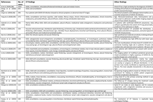
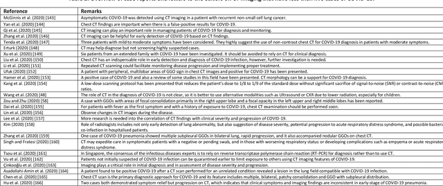
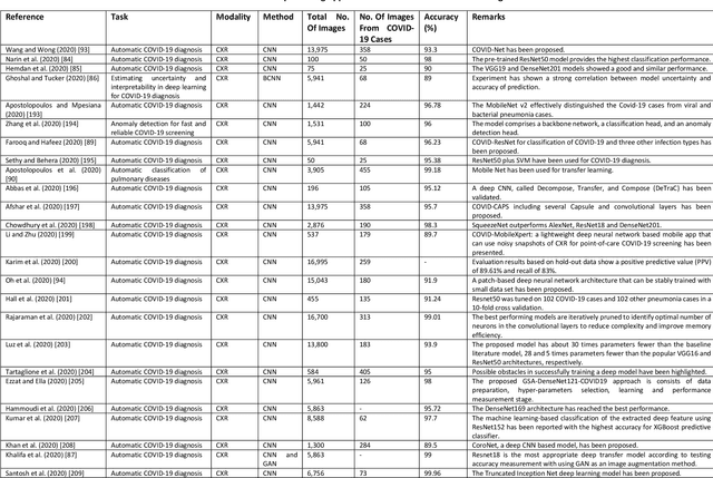
Abstract:Coronavirus disease (COVID-19) is an infectious disease caused by a newly discovered coronavirus. The disease presents with symptoms such as shortness of breath, fever, dry cough, and chronic fatigue, amongst others. Sometimes the symptoms of the disease increase so much they lead to the death of the patients. The disease may be asymptomatic in some patients in the early stages, which can lead to increased transmission of the disease to others. Many studies have tried to use medical imaging for early diagnosis of COVID-19. This study attempts to review papers on automatic methods for medical image analysis and diagnosis of COVID-19. For this purpose, PubMed, Google Scholar, arXiv and medRxiv were searched to find related studies by the end of April 2020, and the essential points of the collected studies were summarised. The contribution of this study is four-fold: 1) to use as a tutorial of the field for both clinicians and technologists, 2) to comprehensively review the characteristics of COVID-19 as presented in medical images, 3) to examine automated artificial intelligence-based approaches for COVID-19 diagnosis based on the accuracy and the method used, 4) to express the research limitations in this field and the methods used to overcome them. COVID-19 reveals signs in medical images can be used for early diagnosis of the disease even in asymptomatic patients. Using automated machine learning-based methods can diagnose the disease with high accuracy from medical images and reduce time, cost and error of diagnostic procedure. It is recommended to collect bulk imaging data from patients in the shortest possible time to improve the performance of COVID-19 automated diagnostic methods.
 Add to Chrome
 Add to Firefox
 Add to Edge