Greg M. Osgood
Exploiting Partial Structural Symmetry For Patient-Specific Image Augmentation in Trauma Interventions
Apr 09, 2018


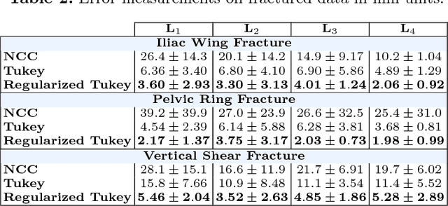
Abstract:In unilateral pelvic fracture reductions, surgeons attempt to reconstruct the bone fragments such that bilateral symmetry in the bony anatomy is restored. We propose to exploit this "structurally symmetric" nature of the pelvic bone, and provide intra-operative image augmentation to assist the surgeon in repairing dislocated fragments. The main challenge is to automatically estimate the desired plane of symmetry within the patient's pre-operative CT. We propose to estimate this plane using a non-linear optimization strategy, by minimizing Tukey's biweight robust estimator, relying on the partial symmetry of the anatomy. Moreover, a regularization term is designed to enforce the similarity of bone density histograms on both sides of this plane, relying on the biological fact that, even if injured, the dislocated bone segments remain within the body. The experimental results demonstrate the performance of the proposed method in estimating this "plane of partial symmetry" using CT images of both healthy and injured anatomy. Examples of unilateral pelvic fractures are used to show how intra-operative X-ray images could be augmented with the forward-projections of the mirrored anatomy, acting as objective road-map for fracture reduction procedures.
 Add to Chrome
 Add to Firefox
 Add to Edge