Geoffrey Jones
Fast Estimation of Haemoglobin Concentration in Tissue Via Wavelet Decomposition
Jun 22, 2017


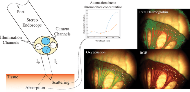
Abstract:Tissue oxygenation and perfusion can be an indicator for organ viability during minimally invasive surgery, for example allowing real-time assessment of tissue perfusion and oxygen saturation. Multispectral imaging is an optical modality that can inspect tissue perfusion in wide field images without contact. In this paper, we present a novel, fast method for using RGB images for MSI, which while limiting the spectral resolution of the modality allows normal laparoscopic systems to be used. We exploit the discrete Haar decomposition to separate individual video frames into low pass and directional coefficients and we utilise a different multispectral estimation technique on each. The increase in speed is achieved by using fast Tikhonov regularisation on the directional coefficients and more accurate Bayesian estimation on the low pass component. The pipeline is implemented using a graphics processing unit (GPU) architecture and achieves a frame rate of approximately 15Hz. We validate the method on animal models and on human data captured using a da Vinci stereo laparoscope.
Inference of Haemoglobin Concentration From Stereo RGB
Jun 22, 2017


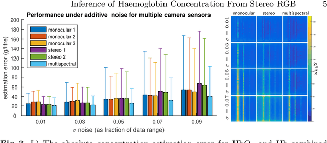
Abstract:Multispectral imaging (MSI) can provide information about tissue oxygenation, perfusion and potentially function during surgery. In this paper we present a novel, near real-time technique for intrinsic measurements of total haemoglobin (THb) and blood oxygenation (SO2) in tissue using only RGB images from a stereo laparoscope. The high degree of spectral overlap between channels makes inference of haemoglobin concentration challenging, non-linear and under constrained. We decompose the problem into two constrained linear sub-problems and show that with Tikhonov regularisation the estimation significantly improves, giving robust estimation of the Thb. We demonstrate by using the co-registered stereo image data from two cameras it is possible to get robust SO2 estimation as well. Our method is closed from, providing computational efficiency even with multiple cameras. The method we present requires only spectral response calibration of each camera, without modification of existing laparoscopic imaging hardware. We validate our technique on synthetic data from Monte Carlo simulation % of light transport through soft tissue containing submerged blood vessels and further, in vivo, on a multispectral porcine data set.
 Add to Chrome
 Add to Firefox
 Add to Edge