Furen Xiao
Parallel Swin Transformer-Enhanced 3D MRI-to-CT Synthesis for MRI-Only Radiotherapy Planning
Feb 05, 2026Abstract:MRI provides superior soft tissue contrast without ionizing radiation; however, the absence of electron density information limits its direct use for dose calculation. As a result, current radiotherapy workflows rely on combined MRI and CT acquisitions, increasing registration uncertainty and procedural complexity. Synthetic CT generation enables MRI only planning but remains challenging due to nonlinear MRI-CT relationships and anatomical variability. We propose Parallel Swin Transformer-Enhanced Med2Transformer, a 3D architecture that integrates convolutional encoding with dual Swin Transformer branches to model both local anatomical detail and long-range contextual dependencies. Multi-scale shifted window attention with hierarchical feature aggregation improves anatomical fidelity. Experiments on public and clinical datasets demonstrate higher image similarity and improved geometric accuracy compared with baseline methods. Dosimetric evaluation shows clinically acceptable performance, with a mean target dose error of 1.69%. Code is available at: https://github.com/mobaidoctor/med2transformer.
Polyp-DDPM: Diffusion-Based Semantic Polyp Synthesis for Enhanced Segmentation
Feb 06, 2024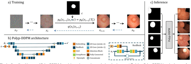
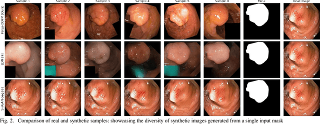
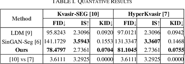
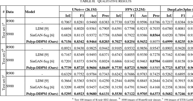
Abstract:This study introduces Polyp-DDPM, a diffusion-based method for generating realistic images of polyps conditioned on masks, aimed at enhancing the segmentation of gastrointestinal (GI) tract polyps. Our approach addresses the challenges of data limitations, high annotation costs, and privacy concerns associated with medical images. By conditioning the diffusion model on segmentation masks-binary masks that represent abnormal areas-Polyp-DDPM outperforms state-of-the-art methods in terms of image quality (achieving a Frechet Inception Distance (FID) score of 78.47, compared to scores above 83.79) and segmentation performance (achieving an Intersection over Union (IoU) of 0.7156, versus less than 0.6694 for synthetic images from baseline models and 0.7067 for real data). Our method generates a high-quality, diverse synthetic dataset for training, thereby enhancing polyp segmentation models to be comparable with real images and offering greater data augmentation capabilities to improve segmentation models. The source code and pretrained weights for Polyp-DDPM are made publicly available at https://github.com/mobaidoctor/polyp-ddpm.
Conditional Diffusion Models for Semantic 3D Medical Image Synthesis
May 29, 2023


Abstract:This paper introduces Med-DDPM, an innovative solution using diffusion models for semantic 3D medical image synthesis, addressing the prevalent issues in medical imaging such as data scarcity, inconsistent acquisition methods, and privacy concerns. Experimental evidence illustrates that diffusion models surpass Generative Adversarial Networks (GANs) in stability and performance, generating high-quality, realistic 3D medical images. The distinct feature of Med-DDPM is its use of semantic conditioning for the diffusion model in 3D image synthesis. By controlling the generation process through pixel-level mask labels, it facilitates the creation of realistic medical images. Empirical evaluations underscore the superior performance of Med-DDPM over GAN techniques in metrics such as accuracy, stability, and versatility. Furthermore, Med-DDPM outperforms traditional augmentation techniques and synthetic GAN images in enhancing the accuracy of segmentation models. It addresses challenges such as insufficient datasets, lack of annotated data, and class imbalance. Noting the limitations of the Frechet inception distance (FID) metric, we introduce a histogram-equalized FID metric for effective performance evaluation. In summary, Med-DDPM, by utilizing diffusion models, signifies a crucial step forward in the domain of high-resolution semantic 3D medical image synthesis, transcending the limitations of GANs and data constraints. This method paves the way for a promising solution in medical imaging, primarily for data augmentation and anonymization, thus contributing significantly to the field.
Deep Learning Based Segmentation of Various Brain Lesions for Radiosurgery
Jul 22, 2020


Abstract:Semantic segmentation of medical images with deep learning models is rapidly developed. In this study, we benchmarked state-of-the-art deep learning segmentation algorithms on our clinical stereotactic radiosurgery dataset, demonstrating the strengths and weaknesses of these algorithms in a fairly practical scenario. In particular, we compared the model performances with respect to their sampling method, model architecture, and the choice of loss functions, identifying the suitable settings for their applications and shedding light on the possible improvements.
Multimodal Volume-Aware Detection and Segmentation for Brain Metastases Radiosurgery
Aug 15, 2019


Abstract:Stereotactic radiosurgery (SRS), which delivers high doses of irradiation in a single or few shots to small targets, has been a standard of care for brain metastases. While very effective, SRS currently requires manually intensive delineation of tumors. In this work, we present a deep learning approach for automated detection and segmentation of brain metastases using multimodal imaging and ensemble neural networks. In order to address small and multiple brain metastases, we further propose a volume-aware Dice loss which optimizes model performance using the information of lesion size. This work surpasses current benchmark levels and demonstrates a reliable AI-assisted system for SRS treatment planning for multiple brain metastases.
A Simple, Fast and Fully Automated Approach for Midline Shift Measurement on Brain Computed Tomography
Mar 02, 2017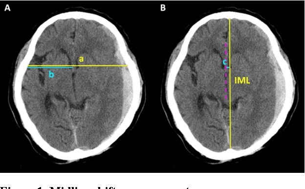
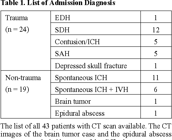
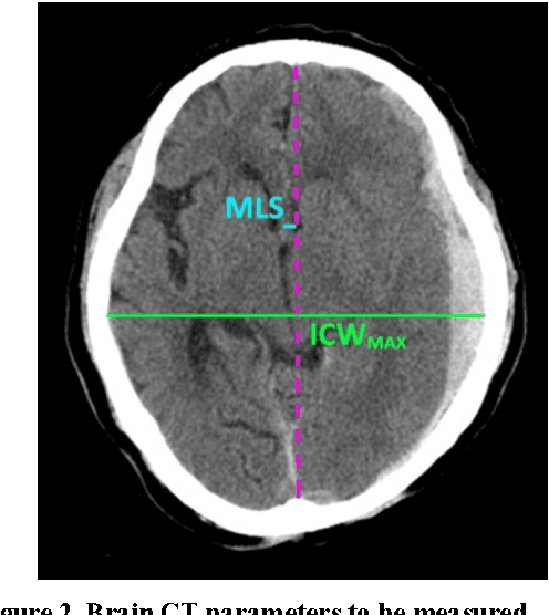
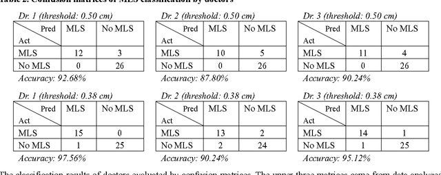
Abstract:Brain CT has become a standard imaging tool for emergent evaluation of brain condition, and measurement of midline shift (MLS) is one of the most important features to address for brain CT assessment. We present a simple method to estimate MLS and propose a new alternative parameter to MLS: the ratio of MLS over the maximal width of intracranial region (MLS/ICWMAX). Three neurosurgeons and our automated system were asked to measure MLS and MLS/ICWMAX in the same sets of axial CT images obtained from 41 patients admitted to ICU under neurosurgical service. A weighted midline (WML) was plotted based on individual pixel intensities, with higher weighted given to the darker portions. The MLS could then be measured as the distance between the WML and ideal midline (IML) near the foramen of Monro. The average processing time to output an automatic MLS measurement was around 10 seconds. Our automated system achieved an overall accuracy of 90.24% when the CT images were calibrated automatically, and performed better when the calibrations of head rotation were done manually (accuracy: 92.68%). MLS/ICWMAX and MLS both gave results in same confusion matrices and produced similar ROC curve results. We demonstrated a simple, fast and accurate automated system of MLS measurement and introduced a new parameter (MLS/ICWMAX) as a good alternative to MLS in terms of estimating the degree of brain deformation, especially when non-DICOM images (e.g. JPEG) are more easily accessed.
 Add to Chrome
 Add to Firefox
 Add to Edge