Fides Regina Schwartz
FPN-IAIA-BL: A Multi-Scale Interpretable Deep Learning Model for Classification of Mass Margins in Digital Mammography
Jun 10, 2024


Abstract:Digital mammography is essential to breast cancer detection, and deep learning offers promising tools for faster and more accurate mammogram analysis. In radiology and other high-stakes environments, uninterpretable ("black box") deep learning models are unsuitable and there is a call in these fields to make interpretable models. Recent work in interpretable computer vision provides transparency to these formerly black boxes by utilizing prototypes for case-based explanations, achieving high accuracy in applications including mammography. However, these models struggle with precise feature localization, reasoning on large portions of an image when only a small part is relevant. This paper addresses this gap by proposing a novel multi-scale interpretable deep learning model for mammographic mass margin classification. Our contribution not only offers an interpretable model with reasoning aligned with radiologist practices, but also provides a general architecture for computer vision with user-configurable prototypes from coarse- to fine-grained prototypes.
Interpretable Mammographic Image Classification using Cased-Based Reasoning and Deep Learning
Jul 12, 2021


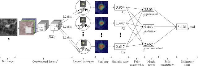
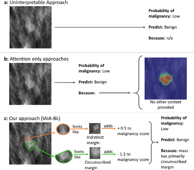
Abstract:When we deploy machine learning models in high-stakes medical settings, we must ensure these models make accurate predictions that are consistent with known medical science. Inherently interpretable networks address this need by explaining the rationale behind each decision while maintaining equal or higher accuracy compared to black-box models. In this work, we present a novel interpretable neural network algorithm that uses case-based reasoning for mammography. Designed to aid a radiologist in their decisions, our network presents both a prediction of malignancy and an explanation of that prediction using known medical features. In order to yield helpful explanations, the network is designed to mimic the reasoning processes of a radiologist: our network first detects the clinically relevant semantic features of each image by comparing each new image with a learned set of prototypical image parts from the training images, then uses those clinical features to predict malignancy. Compared to other methods, our model detects clinical features (mass margins) with equal or higher accuracy, provides a more detailed explanation of its prediction, and is better able to differentiate the classification-relevant parts of the image.
IAIA-BL: A Case-based Interpretable Deep Learning Model for Classification of Mass Lesions in Digital Mammography
Mar 23, 2021


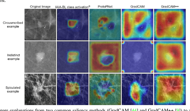
Abstract:Interpretability in machine learning models is important in high-stakes decisions, such as whether to order a biopsy based on a mammographic exam. Mammography poses important challenges that are not present in other computer vision tasks: datasets are small, confounding information is present, and it can be difficult even for a radiologist to decide between watchful waiting and biopsy based on a mammogram alone. In this work, we present a framework for interpretable machine learning-based mammography. In addition to predicting whether a lesion is malignant or benign, our work aims to follow the reasoning processes of radiologists in detecting clinically relevant semantic features of each image, such as the characteristics of the mass margins. The framework includes a novel interpretable neural network algorithm that uses case-based reasoning for mammography. Our algorithm can incorporate a combination of data with whole image labelling and data with pixel-wise annotations, leading to better accuracy and interpretability even with a small number of images. Our interpretable models are able to highlight the classification-relevant parts of the image, whereas other methods highlight healthy tissue and confounding information. Our models are decision aids, rather than decision makers, aimed at better overall human-machine collaboration. We do not observe a loss in mass margin classification accuracy over a black box neural network trained on the same data.
 Add to Chrome
 Add to Firefox
 Add to Edge