Dohyeun Kim
TRk-CNN: Transferable Ranking-CNN for image classification of glaucoma, glaucoma suspect, and normal eyes
May 16, 2019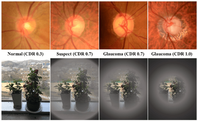


Abstract:In this paper, we proposed Transferable Ranking Convolutional Neural Network (TRk-CNN) that can be effectively applied when the classes of images to be classified show a high correlation with each other. The multi-class classification method based on the softmax function, which is generally used, is not effective in this case because the inter-class relationship is ignored. Although there is a Ranking-CNN that takes into account the ordinal classes, it cannot reflect the inter-class relationship to the final prediction. TRk-CNN, on the other hand, combines the weights of the primitive classification model to reflect the inter-class information to the final classification phase. We evaluated TRk-CNN in glaucoma image dataset that was labeled into three classes: normal, glaucoma suspect, and glaucoma eyes. Based on the literature we surveyed, this study is the first to classify three status of glaucoma fundus image dataset into three different classes. We compared the evaluation results of TRk-CNN with Ranking-CNN (Rk-CNN) and multi-class CNN (MC-CNN) using the DenseNet as the backbone CNN model. As a result, TRk-CNN achieved an average accuracy of 92.96%, specificity of 93.33%, sensitivity for glaucoma suspect of 95.12% and sensitivity for glaucoma of 93.98%. Based on average accuracy, TRk-CNN is 8.04% and 9.54% higher than Rk-CNN and MC-CNN and surprisingly 26.83% higher for sensitivity for suspicious than multi-class CNN. Our TRk-CNN is expected to be effectively applied to the medical image classification problem where the disease state is continuous and increases in the positive class direction.
Tournament Based Ranking CNN for the Cataract grading
Jul 07, 2018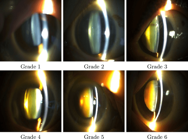
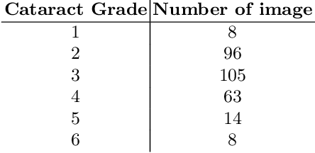
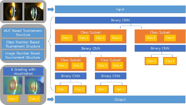

Abstract:Solving the classification problem, unbalanced number of dataset among the classes often causes performance degradation. Especially when some classes dominate the other classes with its large number of datasets, trained model shows low performance in identifying the dominated classes. This is common case when it comes to medical dataset. Because the case with a serious degree is not quite usual, there are imbalance in number of dataset between severe case and normal cases of diseases. Also, there is difficulty in precisely identifying grade of medical data because of vagueness between them. To solve these problems, we propose new architecture of convolutional neural network named Tournament based Ranking CNN which shows remarkable performance gain in identifying dominated classes while trading off very small accuracy loss in dominating classes. Our Approach complemented problems that occur when method of Ranking CNN that aggregates outputs of multiple binary neural network models is applied to medical data. By having tournament structure in aggregating method and using very deep pretrained binary models, our proposed model recorded 68.36% of exact match accuracy, while Ranking CNN recorded 53.40%, pretrained Resnet recorded 56.12% and CNN with linear regression recorded 57.48%. As a result, our proposed method is applied efficiently to cataract grading which have ordinal labels with imbalanced number of data among classes, also can be applied further to medical problems which have similar features to cataract and similar dataset configuration.
2sRanking-CNN: A 2-stage ranking-CNN for diagnosis of glaucoma from fundus images using CAM-extracted ROI as an intermediate input
Jul 04, 2018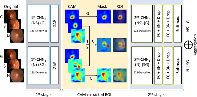

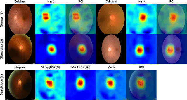

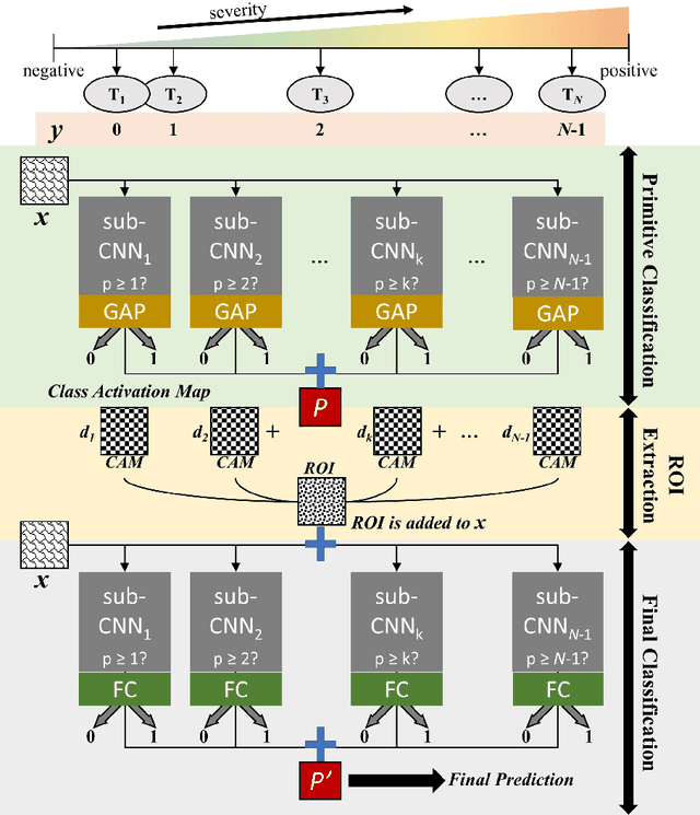
Abstract:Glaucoma is a disease in which the optic nerve is chronically damaged by the elevation of the intra-ocular pressure, resulting in visual field defect. Therefore, it is important to monitor and treat suspected patients before they are confirmed with glaucoma. In this paper, we propose a 2-stage ranking-CNN that classifies fundus images as normal, suspicious, and glaucoma. Furthermore, we propose a method of using the class activation map as a mask filter and combining it with the original fundus image as an intermediate input. Our results have improved the average accuracy by about 10% over the existing 3-class CNN and ranking-CNN, and especially improved the sensitivity of suspicious class by more than 20% over 3-class CNN. In addition, the extracted ROI was also found to overlap with the diagnostic criteria of the physician. The method we propose is expected to be efficiently applied to any medical data where there is a suspicious condition between normal and disease.
Automated diagnosis of pneumothorax using an ensemble of convolutional neural networks with multi-sized chest radiography images
Apr 18, 2018

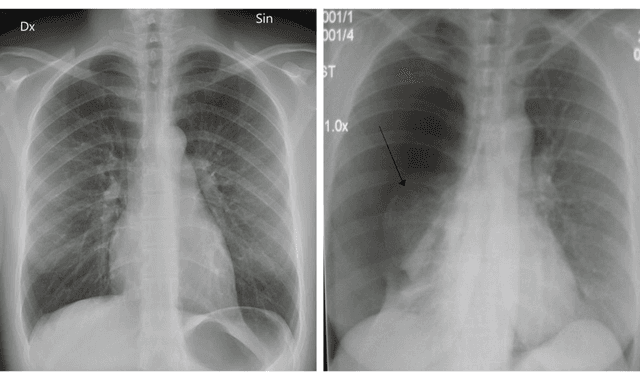

Abstract:Pneumothorax is a relatively common disease, but in some cases, it may be difficult to find with chest radiography. In this paper, we propose a novel method of detecting pneumothorax in chest radiography. We propose an ensemble model of identical convolutional neural networks (CNN) with three different sizes of radiography images. Conventional methods may not properly characterize lost features while resizing large size images into 256 x 256 or 224 x 224 sizes. Our model is evaluated with ChestX-ray dataset which contains over 100,000 chest radiography images. As a result of the experiment, the proposed model showed AUC 0.911, which is the state of the art result in pneumothorax detection. Our method is expected to be effective when applying CNN to large size medical images.
Automated detection of vulnerable plaque in intravascular ultrasound images
Apr 18, 2018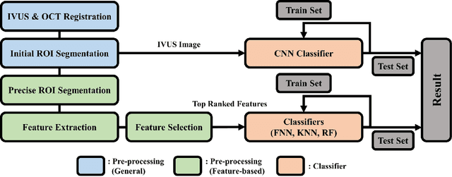
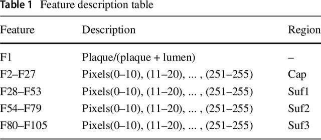
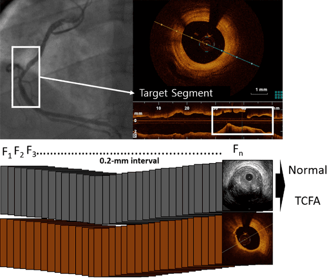
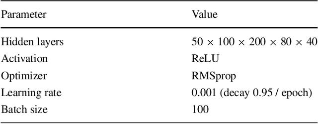
Abstract:Acute Coronary Syndrome (ACS) is a syndrome caused by a decrease in blood flow in the coronary arteries. The ACS is usually related to coronary thrombosis and is primarily caused by plaque rupture followed by plaque erosion and calcified nodule. Thin-cap fibroatheroma (TCFA) is known to be the most similar lesion morphologically to a plaque rupture. In this paper, we propose methods to classify TCFA using various machine learning classifiers including Feed-forward Neural Network (FNN), K-Nearest Neighbor (KNN), Random Forest (RF) and Convolutional Neural Network (CNN) to figure out a classifier that shows optimal TCFA classification accuracy. In addition, we suggest pixel range based feature extraction method to extract the ratio of pixels in the different region of interests to reflect the physician's TCFA discrimination criteria. A total of 12,325 IVUS images were labeled with corresponding OCT images to train and evaluate the classifiers. We achieved 0.884, 0.890, 0.878 and 0.933 Area Under the ROC Curve (AUC) in the order of using FNN, KNN, RF and CNN classifier. As a result, the CNN classifier performed best and the top 10 features of the feature-based classifiers (FNN, KNN, RF) were found to be similar to the physician's TCFA diagnostic criteria.
ECG arrhythmia classification using a 2-D convolutional neural network
Apr 18, 2018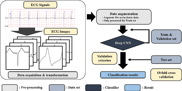


Abstract:In this paper, we propose an effective electrocardiogram (ECG) arrhythmia classification method using a deep two-dimensional convolutional neural network (CNN) which recently shows outstanding performance in the field of pattern recognition. Every ECG beat was transformed into a two-dimensional grayscale image as an input data for the CNN classifier. Optimization of the proposed CNN classifier includes various deep learning techniques such as batch normalization, data augmentation, Xavier initialization, and dropout. In addition, we compared our proposed classifier with two well-known CNN models; AlexNet and VGGNet. ECG recordings from the MIT-BIH arrhythmia database were used for the evaluation of the classifier. As a result, our classifier achieved 99.05% average accuracy with 97.85% average sensitivity. To precisely validate our CNN classifier, 10-fold cross-validation was performed at the evaluation which involves every ECG recording as a test data. Our experimental results have successfully validated that the proposed CNN classifier with the transformed ECG images can achieve excellent classification accuracy without any manual pre-processing of the ECG signals such as noise filtering, feature extraction, and feature reduction.
 Add to Chrome
 Add to Firefox
 Add to Edge