Dina Sheyfer
Optimizing Coded-Apertures for Depth-Resolved Diffraction
May 21, 2024


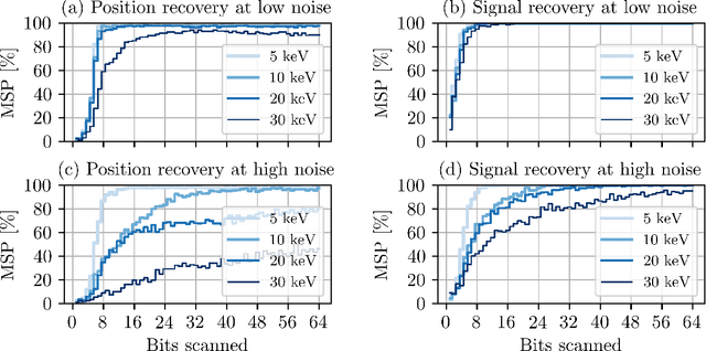
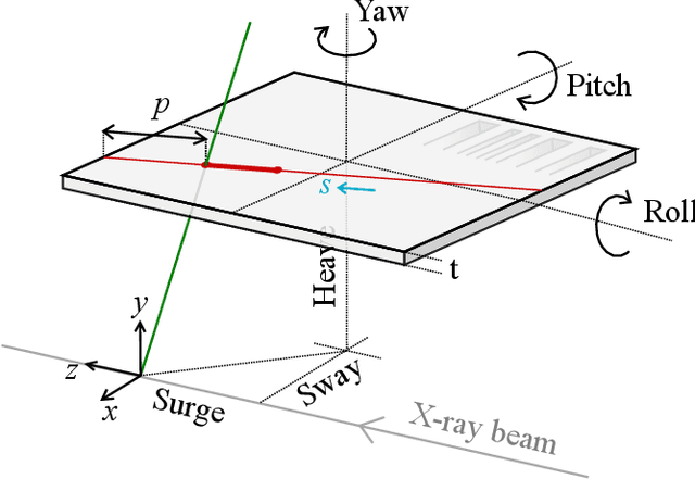
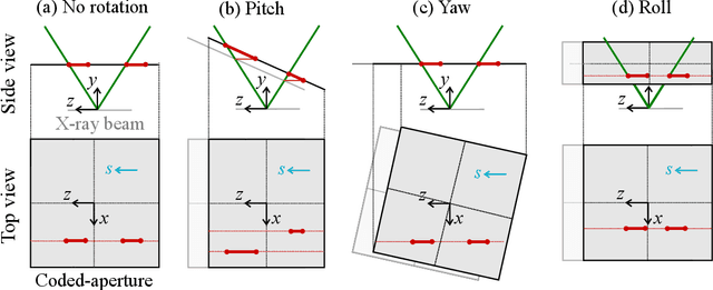
Abstract:Coded apertures, traditionally employed in x-ray astronomy for imaging celestial objects, are now being adapted for micro-scale applications, particularly in studying microscopic specimens with synchrotron light diffraction. In this paper, we focus on micro-coded aperture imaging and its capacity to accomplish depth-resolved micro-diffraction analysis within crystalline specimens. We study aperture specifications and scanning parameters by assessing characteristics like size, thickness, and patterns. Numerical experiments assist in assessing their impact on reconstruction quality. Empirical data from a Laue diffraction microscope at a synchrotron undulator beamline supports our findings. Overall, our results offer key insights for optimizing aperture design in advancing micro-scale diffraction imaging at synchrotrons. This study contributes insights to this expanding field and suggests significant advancements, especially when coupled with the enhanced flux anticipated from the global upgrades of synchrotron sources.
Dark-Field X-Ray Microscopy with Structured Illumination for Three-Dimensional Imaging
May 21, 2024


Abstract:We introduce a structured illumination technique for dark-field x-ray microscopy optimized for three-dimensional imaging of ordered materials at sub-micrometer length scales. Our method utilizes a coded aperture to spatially modulate the incident x-ray beam on the sample, enabling the reconstruction of the sample's 3D structure from images captured at various aperture positions. Unlike common volumetric imaging techniques such as tomography, our approach casts a scanning x-ray silhouette of a coded aperture for depth resolution along the axis of diffraction, eliminating any need for sample rotation or rastering, leading to a highly stable imaging modality. This modification provides robustness against geometric uncertainties during data acquisition, particularly for achieving sub-micrometer resolutions where geometric uncertainties typically limit resolution. We introduce the image reconstruction model and validate our results with experimental data on an isolated twin domain within a bulk single crystal of an iron pnictide obtained using a dark-field x-ray microscope. This timely advancement aligns with the enhanced brightness upgrade of the world's synchrotron radiation facilities, opening unprecedented opportunities in imaging.
Machine learning for interpreting coherent X-ray speckle patterns
Nov 15, 2022Abstract:Speckle patterns produced by coherent X-ray have a close relationship with the internal structure of materials but quantitative inversion of the relationship to determine structure from images is challenging. Here, we investigate the link between coherent X-ray speckle patterns and sample structures using a model 2D disk system and explore the ability of machine learning to learn aspects of the relationship. Specifically, we train a deep neural network to classify the coherent X-ray speckle pattern images according to the disk number density in the corresponding structure. It is demonstrated that the classification system is accurate for both non-disperse and disperse size distributions.
Digital autofocusing of a coded-aperture Laue diffraction microscope
Aug 15, 2022


Abstract:To provide optimal depth resolution with a coded-aperture Laue diffraction microscope, an accurate position of the coded-aperture and its scanning geometry need to be known. However, finding the geometry by trial and error is a time-consuming and often challenging process because of the large number of parameters involved. In this paper, we propose an optimization approach to automate the focusing process after data is collected. We demonstrate the robustness and efficiency of the proposed approach with experimental data taken at a synchrotron facility.
Depth-resolved Laue microdiffraction with coded-apertures
Mar 03, 2022


Abstract:We introduce a rapid data acquisition and reconstruction method to image the crystalline structure of materials and associated strain and orientations at micrometer resolution using Laue diffraction. Our method relies on scanning a coded-aperture across the diffracted x-ray beams from a broadband illumination, and a reconstruction algorithm to resolve Laue microdiffraction patterns as a function of depth along the incident illumination path. This method provides a rapid access to full diffraction information at sub-micrometer volume elements in bulk materials. Here we present the theory as well as the experimental validation of this imaging approach.
 Add to Chrome
 Add to Firefox
 Add to Edge