Charudatta Phatak
High resolution functional imaging through Lorentz transmission electron microscopy and differentiable programming
Dec 07, 2020


Abstract:Lorentz transmission electron microscopy is a unique characterization technique that enables the simultaneous imaging of both the microstructure and functional properties of materials at high spatial resolution. The quantitative information such as magnetization and electric potentials is carried by the phase of the electron wave, and is lost during imaging. In order to understand the local interactions and develop structure-property relationships, it is necessary to retrieve the complete wavefunction of the electron wave, which requires solving for the phase shift of the electrons (phase retrieval). Here we have developed a method based on differentiable programming to solve the inverse problem of phase retrieval, using a series of defocused microscope images. We show that our method is robust and can outperform widely used \textit{transport of intensity equation} in terms of spatial resolution and accuracy of the retrieved phase under same electron dose conditions. Furthermore, our method shares the same basic structure as advanced machine learning algorithms, and is easily adaptable to various other forms of phase retrieval in electron microscopy.
U-SLADS: Unsupervised Learning Approach for Dynamic Dendrite Sampling
Jul 06, 2018
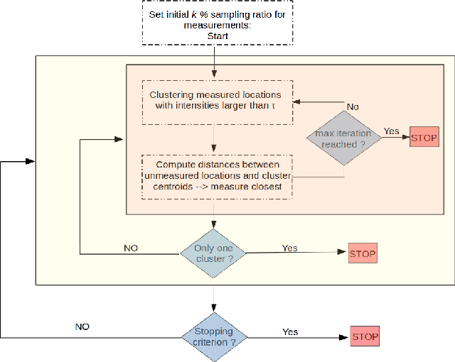
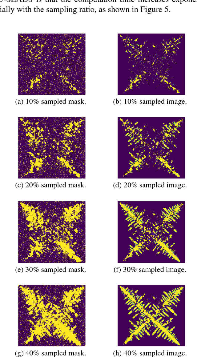
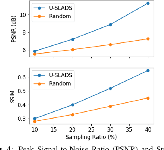
Abstract:Novel data acquisition schemes have been an emerging need for scanning microscopy based imaging techniques to reduce the time in data acquisition and to minimize probing radiation in sample exposure. Varies sparse sampling schemes have been studied and are ideally suited for such applications where the images can be reconstructed from a sparse set of measurements. Dynamic sparse sampling methods, particularly supervised learning based iterative sampling algorithms, have shown promising results for sampling pixel locations on the edges or boundaries during imaging. However, dynamic sampling for imaging skeleton-like objects such as metal dendrites remains difficult. Here, we address a new unsupervised learning approach using Hierarchical Gaussian Mixture Mod- els (HGMM) to dynamically sample metal dendrites. This technique is very useful if the users are interested in fast imaging the primary and secondary arms of metal dendrites in solidification process in materials science.
Reduced Electron Exposure for Energy-Dispersive Spectroscopy using Dynamic Sampling
Jun 27, 2017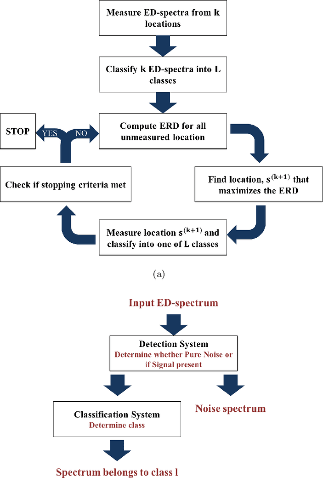
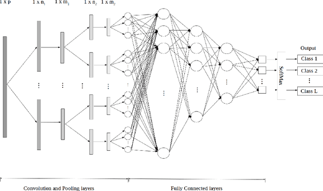
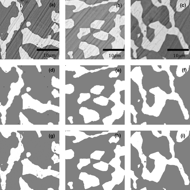
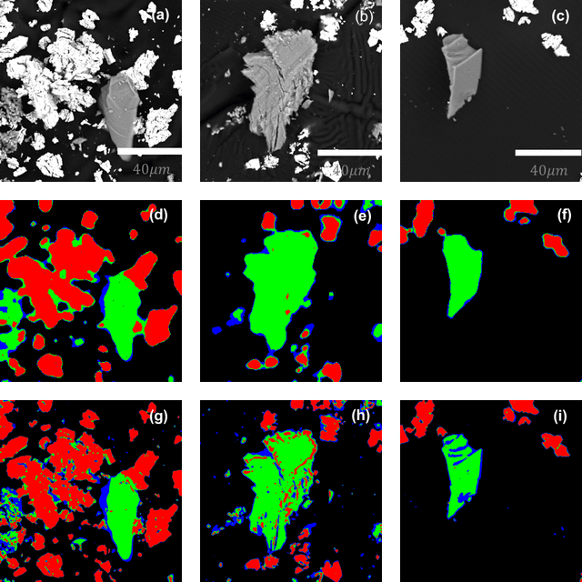
Abstract:Analytical electron microscopy and spectroscopy of biological specimens, polymers, and other beam sensitive materials has been a challenging area due to irradiation damage. There is a pressing need to develop novel imaging and spectroscopic imaging methods that will minimize such sample damage as well as reduce the data acquisition time. The latter is useful for high-throughput analysis of materials structure and chemistry. In this work, we present a novel machine learning based method for dynamic sparse sampling of EDS data using a scanning electron microscope. Our method, based on the supervised learning approach for dynamic sampling algorithm and neural networks based classification of EDS data, allows a dramatic reduction in the total sampling of up to 90%, while maintaining the fidelity of the reconstructed elemental maps and spectroscopic data. We believe this approach will enable imaging and elemental mapping of materials that would otherwise be inaccessible to these analysis techniques.
 Add to Chrome
 Add to Firefox
 Add to Edge