Cesar Caballero-Gaudes
TV-based Deep 3D Self Super-Resolution for fMRI
Oct 05, 2024


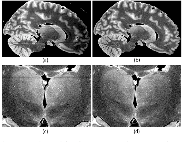
Abstract:While functional Magnetic Resonance Imaging (fMRI) offers valuable insights into cognitive processes, its inherent spatial limitations pose challenges for detailed analysis of the fine-grained functional architecture of the brain. More specifically, MRI scanner and sequence specifications impose a trade-off between temporal resolution, spatial resolution, signal-to-noise ratio, and scan time. Deep Learning (DL) Super-Resolution (SR) methods have emerged as a promising solution to enhance fMRI resolution, generating high-resolution (HR) images from low-resolution (LR) images typically acquired with lower scanning times. However, most existing SR approaches depend on supervised DL techniques, which require training ground truth (GT) HR data, which is often difficult to acquire and simultaneously sets a bound for how far SR can go. In this paper, we introduce a novel self-supervised DL SR model that combines a DL network with an analytical approach and Total Variation (TV) regularization. Our method eliminates the need for external GT images, achieving competitive performance compared to supervised DL techniques and preserving the functional maps.
A probabilistic atlas of the human thalamic nuclei combining ex vivo MRI and histology
Jun 22, 2018


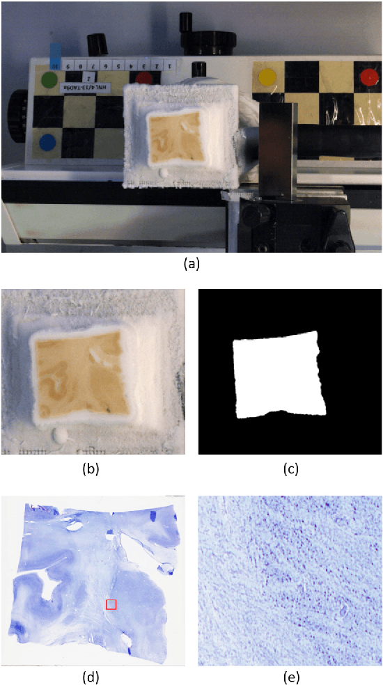
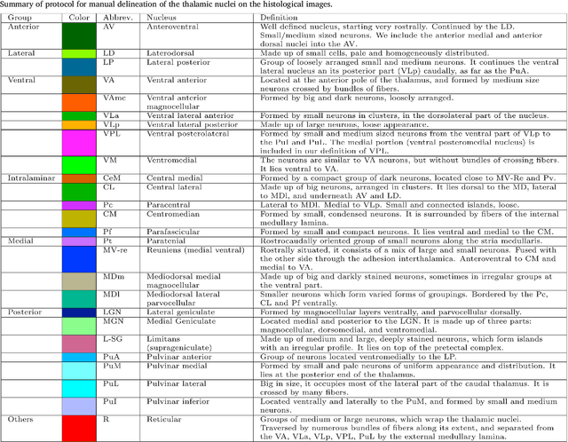
Abstract:The human thalamus is a brain structure that comprises numerous, highly specific nuclei. Since these nuclei are known to have different functions and to be connected to different areas of the cerebral cortex, it is of great interest for the neuroimaging community to study their volume, shape and connectivity in vivo with MRI. In this study, we present a probabilistic atlas of the thalamic nuclei built using ex vivo brain MRI scans and histological data, as well as the application of the atlas to in vivo MRI segmentation. The atlas was built using manual delineation of 26 thalamic nuclei on the serial histology of 12 whole thalami from six autopsy samples, combined with manual segmentations of the whole thalamus and surrounding structures (caudate, putamen, hippocampus, etc.) made on in vivo brain MR data from 39 subjects. The 3D structure of the histological data and corresponding manual segmentations was recovered using the ex vivo MRI as reference frame, and stacks of blockface photographs acquired during the sectioning as intermediate target. The atlas, which was encoded as an adaptive tetrahedral mesh, shows a good agreement with with previous histological studies of the thalamus in terms of volumes of representative nuclei. When applied to segmentation of in vivo scans using Bayesian inference, the atlas shows excellent test-retest reliability, robustness to changes in input MRI contrast, and ability to detect differential thalamic effects in subjects with Alzheimer's disease. The probabilistic atlas and companion segmentation tool are publicly available as part of the neuroimaging package FreeSurfer.
 Add to Chrome
 Add to Firefox
 Add to Edge