Asim Naveed
AD-Net: Attention-based dilated convolutional residual network with guided decoder for robust skin lesion segmentation
Sep 09, 2024Abstract:In computer-aided diagnosis tools employed for skin cancer treatment and early diagnosis, skin lesion segmentation is important. However, achieving precise segmentation is challenging due to inherent variations in appearance, contrast, texture, and blurry lesion boundaries. This research presents a robust approach utilizing a dilated convolutional residual network, which incorporates an attention-based spatial feature enhancement block (ASFEB) and employs a guided decoder strategy. In each dilated convolutional residual block, dilated convolution is employed to broaden the receptive field with varying dilation rates. To improve the spatial feature information of the encoder, we employed an attention-based spatial feature enhancement block in the skip connections. The ASFEB in our proposed method combines feature maps obtained from average and maximum-pooling operations. These combined features are then weighted using the active outcome of global average pooling and convolution operations. Additionally, we have incorporated a guided decoder strategy, where each decoder block is optimized using an individual loss function to enhance the feature learning process in the proposed AD-Net. The proposed AD-Net presents a significant benefit by necessitating fewer model parameters compared to its peer methods. This reduction in parameters directly impacts the number of labeled data required for training, facilitating faster convergence during the training process. The effectiveness of the proposed AD-Net was evaluated using four public benchmark datasets. We conducted a Wilcoxon signed-rank test to verify the efficiency of the AD-Net. The outcomes suggest that our method surpasses other cutting-edge methods in performance, even without the implementation of data augmentation strategies.
TBConvL-Net: A Hybrid Deep Learning Architecture for Robust Medical Image Segmentation
Sep 05, 2024


Abstract:Deep learning has shown great potential for automated medical image segmentation to improve the precision and speed of disease diagnostics. However, the task presents significant difficulties due to variations in the scale, shape, texture, and contrast of the pathologies. Traditional convolutional neural network (CNN) models have certain limitations when it comes to effectively modelling multiscale context information and facilitating information interaction between skip connections across levels. To overcome these limitations, a novel deep learning architecture is introduced for medical image segmentation, taking advantage of CNNs and vision transformers. Our proposed model, named TBConvL-Net, involves a hybrid network that combines the local features of a CNN encoder-decoder architecture with long-range and temporal dependencies using biconvolutional long-short-term memory (LSTM) networks and vision transformers (ViT). This enables the model to capture contextual channel relationships in the data and account for the uncertainty of segmentation over time. Additionally, we introduce a novel composite loss function that considers both the segmentation robustness and the boundary agreement of the predicted output with the gold standard. Our proposed model shows consistent improvement over the state of the art on ten publicly available datasets of seven different medical imaging modalities.
LMBiS-Net: A Lightweight Multipath Bidirectional Skip Connection based CNN for Retinal Blood Vessel Segmentation
Sep 10, 2023Abstract:Blinding eye diseases are often correlated with altered retinal morphology, which can be clinically identified by segmenting retinal structures in fundus images. However, current methodologies often fall short in accurately segmenting delicate vessels. Although deep learning has shown promise in medical image segmentation, its reliance on repeated convolution and pooling operations can hinder the representation of edge information, ultimately limiting overall segmentation accuracy. In this paper, we propose a lightweight pixel-level CNN named LMBiS-Net for the segmentation of retinal vessels with an exceptionally low number of learnable parameters \textbf{(only 0.172 M)}. The network used multipath feature extraction blocks and incorporates bidirectional skip connections for the information flow between the encoder and decoder. Additionally, we have optimized the efficiency of the model by carefully selecting the number of filters to avoid filter overlap. This optimization significantly reduces training time and enhances computational efficiency. To assess the robustness and generalizability of LMBiS-Net, we performed comprehensive evaluations on various aspects of retinal images. Specifically, the model was subjected to rigorous tests to accurately segment retinal vessels, which play a vital role in ophthalmological diagnosis and treatment. By focusing on the retinal blood vessels, we were able to thoroughly analyze the performance and effectiveness of the LMBiS-Net model. The results of our tests demonstrate that LMBiS-Net is not only robust and generalizable but also capable of maintaining high levels of segmentation accuracy. These characteristics highlight the potential of LMBiS-Net as an efficient tool for high-speed and accurate segmentation of retinal images in various clinical applications.
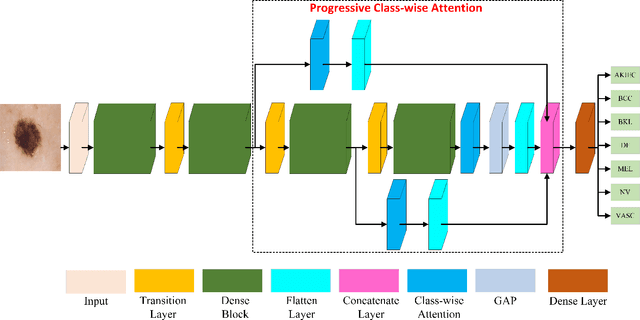
Progressive Class-Wise Attention (PCA) Approach for Diagnosing Skin Lesions
Jun 11, 2023


Abstract:Skin cancer holds the highest incidence rate among all cancers globally. The importance of early detection cannot be overstated, as late-stage cases can be lethal. Classifying skin lesions, however, presents several challenges due to the many variations they can exhibit, such as differences in colour, shape, and size, significant variation within the same class, and notable similarities between different classes. This paper introduces a novel class-wise attention technique that equally regards each class while unearthing more specific details about skin lesions. This attention mechanism is progressively used to amalgamate discriminative feature details from multiple scales. The introduced technique demonstrated impressive performance, surpassing more than 15 cutting-edge methods including the winners of HAM1000 and ISIC 2019 leaderboards. It achieved an impressive accuracy rate of 97.40% on the HAM10000 dataset and 94.9% on the ISIC 2019 dataset.
 Add to Chrome
 Add to Firefox
 Add to Edge