Zeina Al Masry
FEMTO-ST
A new methodology to predict the oncotype scores based on clinico-pathological data with similar tumor profiles
Mar 13, 2023Abstract:Introduction: The Oncotype DX (ODX) test is a commercially available molecular test for breast cancer assay that provides prognostic and predictive breast cancer recurrence information for hormone positive, HER2-negative patients. The aim of this study is to propose a novel methodology to assist physicians in their decision-making. Methods: A retrospective study between 2012 and 2020 with 333 cases that underwent an ODX assay from three hospitals in Bourgogne Franche-Comt{\'e} was conducted. Clinical and pathological reports were used to collect the data. A methodology based on distributional random forest was developed using 9 clinico-pathological characteristics. This methodology can be used particularly to identify the patients of the training cohort that share similarities with the new patient and to predict an estimate of the distribution of the ODX score. Results: The mean age of participants id 56.9 years old. We have correctly classified 92% of patients in low risk and 40.2% of patients in high risk. The overall accuracy is 79.3%. The proportion of low risk correct predicted value (PPV) is 82%. The percentage of high risk correct predicted value (NPV) is approximately 62.3%. The F1-score and the Area Under Curve (AUC) are of 0.87 and 0.759, respectively. Conclusion: The proposed methodology makes it possible to predict the distribution of the ODX score for a patient and provides an explanation of the predicted score. The use of the methodology with the pathologist's expertise on the different histological and immunohistochemical characteristics has a clinical impact to help oncologist in decision-making regarding breast cancer therapy.
A deep learning pipeline for breast cancer ki-67 proliferation index scoring
Mar 14, 2022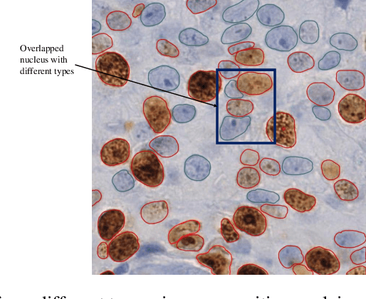

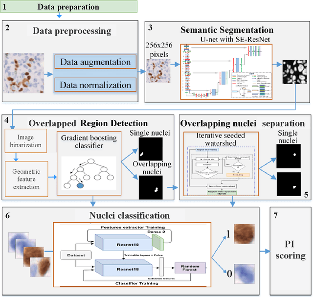

Abstract:The Ki-67 proliferation index is an essential biomarker that helps pathologists to diagnose and select appropriate treatments. However, automatic evaluation of Ki-67 is difficult due to nuclei overlapping and complex variations in their properties. This paper proposes an integrated pipeline for accurate automatic counting of Ki-67, where the impact of nuclei separation techniques is highlighted. First, semantic segmentation is performed by combining the Squeez and Excitation Resnet and Unet algorithms to extract nuclei from the background. The extracted nuclei are then divided into overlapped and non-overlapped regions based on eight geometric and statistical features. A marker-based Watershed algorithm is subsequently proposed and applied only to the overlapped regions to separate nuclei. Finally, deep features are extracted from each nucleus patch using Resnet18 and classified into positive or negative by a random forest classifier. The proposed pipeline's performance is validated on a dataset from the Department of Pathology at H\^opital Nord Franche-Comt\'e hospital.
A CNN-based methodology for breast cancer diagnosis using thermal images
Oct 30, 2019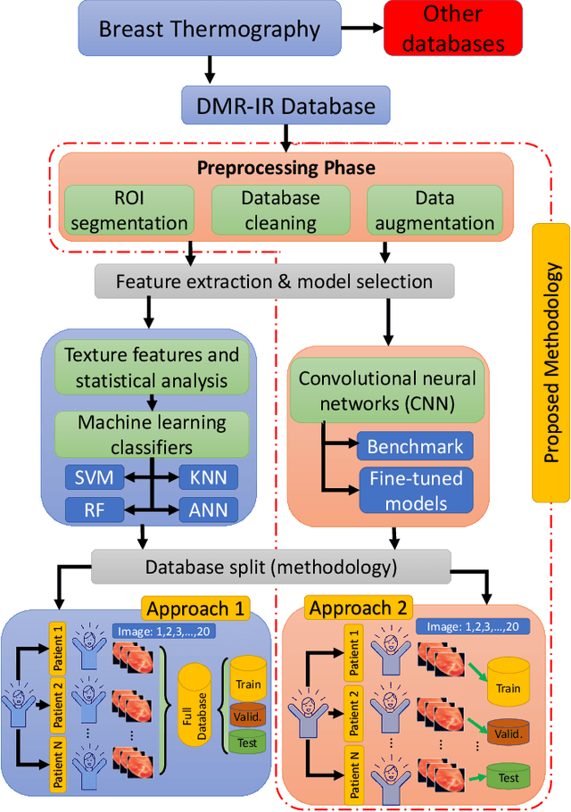
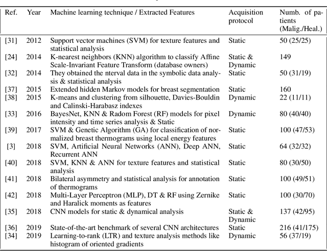
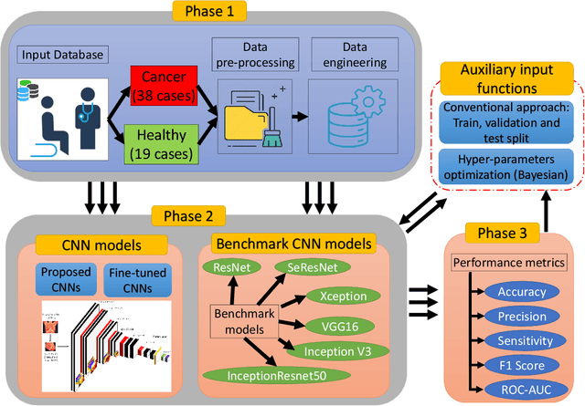
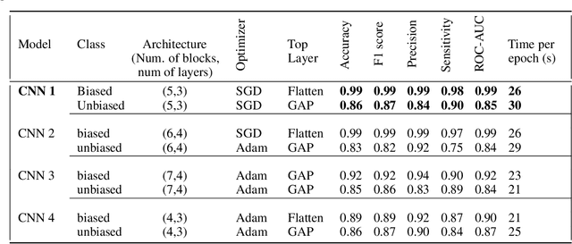
Abstract:Micro Abstract: A recent study from GLOBOCAN disclosed that during 2018 two million women worldwide had been diagnosed from breast cancer. This study presents a computer-aided diagnosis system based on convolutional neural networks as an alternative diagnosis methodology for breast cancer diagnosis with thermal images. Experimental results showed that lower false-positives and false-negatives classification rates are obtained when data pre-processing and data augmentation techniques are implemented in these thermal images. Background: There are many types of breast cancer screening techniques such as, mammography, magnetic resonance imaging, ultrasound and blood sample tests, which require either, expensive devices or personal qualified. Currently, some countries still lack access to these main screening techniques due to economic, social or cultural issues. The objective of this study is to demonstrate that computer-aided diagnosis(CAD) systems based on convolutional neural networks (CNN) are faster, reliable and robust than other techniques. Methods: We performed a study of the influence of data pre-processing, data augmentation and database size versus a proposed set of CNN models. Furthermore, we developed a CNN hyper-parameters fine-tuning optimization algorithm using a tree parzen estimator. Results: Among the 57 patients database, our CNN models obtained a higher accuracy (92\%) and F1-score (92\%) that outperforms several state-of-the-art architectures such as ResNet50, SeResNet50 and Inception. Also, we demonstrated that a CNN model that implements data-augmentation techniques reach identical performance metrics in comparison with a CNN that uses a database up to 50\% bigger. Conclusion: This study highlights the benefits of data augmentation and CNNs in thermal breast images. Also, it measures the influence of the database size in the performance of CNNs.
 Add to Chrome
 Add to Firefox
 Add to Edge