Yong-Ping Zheng
SA$^{2}$Net: Scale-Adaptive Structure-Affinity Transformation for Spine Segmentation from Ultrasound Volume Projection Imaging
Oct 30, 2025Abstract:Spine segmentation, based on ultrasound volume projection imaging (VPI), plays a vital role for intelligent scoliosis diagnosis in clinical applications. However, this task faces several significant challenges. Firstly, the global contextual knowledge of spines may not be well-learned if we neglect the high spatial correlation of different bone features. Secondly, the spine bones contain rich structural knowledge regarding their shapes and positions, which deserves to be encoded into the segmentation process. To address these challenges, we propose a novel scale-adaptive structure-aware network (SA$^{2}$Net) for effective spine segmentation. First, we propose a scale-adaptive complementary strategy to learn the cross-dimensional long-distance correlation features for spinal images. Second, motivated by the consistency between multi-head self-attention in Transformers and semantic level affinity, we propose structure-affinity transformation to transform semantic features with class-specific affinity and combine it with a Transformer decoder for structure-aware reasoning. In addition, we adopt a feature mixing loss aggregation method to enhance model training. This method improves the robustness and accuracy of the segmentation process. The experimental results demonstrate that our SA$^{2}$Net achieves superior segmentation performance compared to other state-of-the-art methods. Moreover, the adaptability of SA$^{2}$Net to various backbones enhances its potential as a promising tool for advanced scoliosis diagnosis using intelligent spinal image analysis. The code and experimental demo are available at https://github.com/taetiseo09/SA2Net.
Automatic Ultrasound Curve Angle Measurement via Affinity Clustering for Adolescent Idiopathic Scoliosis Evaluation
May 07, 2024


Abstract:The current clinical gold standard for evaluating adolescent idiopathic scoliosis (AIS) is X-ray radiography, using Cobb angle measurement. However, the frequent monitoring of the AIS progression using X-rays poses a challenge due to the cumulative radiation exposure. Although 3D ultrasound has been validated as a reliable and radiation-free alternative for scoliosis assessment, the process of measuring spinal curvature is still carried out manually. Consequently, there is a considerable demand for a fully automatic system that can locate bony landmarks and perform angle measurements. To this end, we introduce an estimation model for automatic ultrasound curve angle (UCA) measurement. The model employs a dual-branch network to detect candidate landmarks and perform vertebra segmentation on ultrasound coronal images. An affinity clustering strategy is utilized within the vertebral segmentation area to illustrate the affinity relationship between candidate landmarks. Subsequently, we can efficiently perform line delineation from a clustered affinity map for UCA measurement. As our method is specifically designed for UCA calculation, this method outperforms other state-of-the-art methods for landmark and line detection tasks. The high correlation between the automatic UCA and Cobb angle (R$^2$=0.858) suggests that our proposed method can potentially replace manual UCA measurement in ultrasound scoliosis assessment.
Bone Feature Segmentation in Ultrasound Spine Image with Robustness to Speckle and Regular Occlusion Noise
Oct 08, 2020


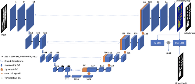
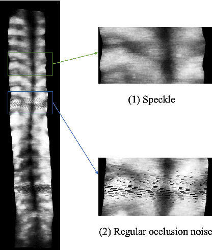
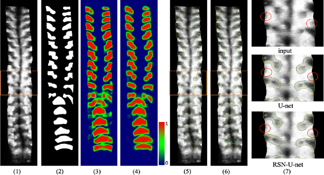
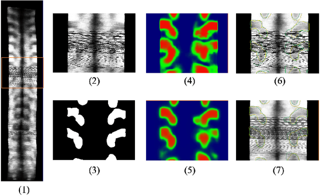
Abstract:3D ultrasound imaging shows great promise for scoliosis diagnosis thanks to its low-costing, radiation-free and real-time characteristics. The key to accessing scoliosis by ultrasound imaging is to accurately segment the bone area and measure the scoliosis degree based on the symmetry of the bone features. The ultrasound images tend to contain many speckles and regular occlusion noise which is difficult, tedious and time-consuming for experts to find out the bony feature. In this paper, we propose a robust bone feature segmentation method based on the U-net structure for ultrasound spine Volume Projection Imaging (VPI) images. The proposed segmentation method introduces a total variance loss to reduce the sensitivity of the model to small-scale and regular occlusion noise. The proposed approach improves 2.3% of Dice score and 1% of AUC score as compared with the u-net model and shows high robustness to speckle and regular occlusion noise.
 Add to Chrome
 Add to Firefox
 Add to Edge