Timothy Tin-Yan Lee
Morphological-consistent Diffusion Network for Ultrasound Coronal Image Enhancement
Sep 25, 2024


Abstract:Ultrasound curve angle (UCA) measurement provides a radiation-free and reliable evaluation for scoliosis based on ultrasound imaging. However, degraded image quality, especially in difficult-to-image patients, can prevent clinical experts from making confident measurements, even leading to misdiagnosis. In this paper, we propose a multi-stage image enhancement framework that models high-quality image distribution via a diffusion-based model. Specifically, we integrate the underlying morphological information from images taken at different depths of the 3D volume to calibrate the reverse process toward high-quality and high-fidelity image generation. This is achieved through a fusion operation with a learnable tuner module that learns the multi-to-one mapping from multi-depth to high-quality images. Moreover, the separate learning of the high-quality image distribution and the spinal features guarantees the preservation of consistent spinal pose descriptions in the generated images, which is crucial in evaluating spinal deformities. Remarkably, our proposed enhancement algorithm significantly outperforms other enhancement-based methods on ultrasound images in terms of image quality. Ultimately, we conduct the intra-rater and inter-rater measurements of UCA and higher ICC (0.91 and 0.89 for thoracic and lumbar angles) on enhanced images, indicating our method facilitates the measurement of ultrasound curve angles and offers promising prospects for automated scoliosis diagnosis.
Automatic Ultrasound Curve Angle Measurement via Affinity Clustering for Adolescent Idiopathic Scoliosis Evaluation
May 07, 2024


Abstract:The current clinical gold standard for evaluating adolescent idiopathic scoliosis (AIS) is X-ray radiography, using Cobb angle measurement. However, the frequent monitoring of the AIS progression using X-rays poses a challenge due to the cumulative radiation exposure. Although 3D ultrasound has been validated as a reliable and radiation-free alternative for scoliosis assessment, the process of measuring spinal curvature is still carried out manually. Consequently, there is a considerable demand for a fully automatic system that can locate bony landmarks and perform angle measurements. To this end, we introduce an estimation model for automatic ultrasound curve angle (UCA) measurement. The model employs a dual-branch network to detect candidate landmarks and perform vertebra segmentation on ultrasound coronal images. An affinity clustering strategy is utilized within the vertebral segmentation area to illustrate the affinity relationship between candidate landmarks. Subsequently, we can efficiently perform line delineation from a clustered affinity map for UCA measurement. As our method is specifically designed for UCA calculation, this method outperforms other state-of-the-art methods for landmark and line detection tasks. The high correlation between the automatic UCA and Cobb angle (R$^2$=0.858) suggests that our proposed method can potentially replace manual UCA measurement in ultrasound scoliosis assessment.
Reliability of Robotic Ultrasound Scanning for Scoliosis Assessment in Comparison with Manual Scanning
May 07, 2022


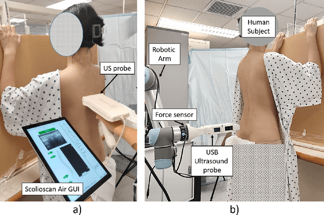
Abstract:Background: Ultrasound (US) imaging for scoliosis assessment is challenging for a non-experienced operator. The robotic scanning was developed to follow a spinal curvature with deep learning and apply consistent forces to the patient' back. Methods: 23 scoliosis patients were scanned with US devices both, robotically and manually. Two human raters measured each subject's spinous process angles (SPA) on robotic and manual coronal images. Results: The robotic method showed high intra- (ICC > 0.85) and inter-rater (ICC > 0.77) reliabilities. Compared with the manual method, the robotic approach showed no significant difference (p < 0.05) when measuring coronal deformity angles. The MAD for intra-rater analysis lies within an acceptable range from 0 deg to 5 deg for a minimum of 86% and a maximum 97% of a total number of the measured angles. Conclusions: This study demonstrated that scoliosis deformity angles measured on ultrasound images obtained with robotic scanning are comparable to those obtained by manual scanning.
 Add to Chrome
 Add to Firefox
 Add to Edge